Floor Couvreur, Elke Loos, Christian Desloovere, Nicolas Verhaert
{"title":"耳内镜在慢性耳显微手术中检测残留胆脂瘤的功效","authors":"Floor Couvreur, Elke Loos, Christian Desloovere, Nicolas Verhaert","doi":"10.5152/iao.2024.231122","DOIUrl":null,"url":null,"abstract":"<p><p>The aim of this article is to determine the efficacy of otoendoscopy during microscopic cholesteatoma surgery on residual cholesteatoma rates postoperatively. The medical records of patients (aged 4-90) with primary acquired cholesteatoma who underwent microscopic cholesteatoma surgery (exclusively transcanal approach or canal wall-up tympano-mastoidectomy) with subsequent otoendoscopic examination (80 ears) for intraoperative cholesteatoma residues were retrospectively reviewed. All cases with mixed microscopic/endoscopic, fully endoscopic, or fully microscopic dissection were excluded, as well as cases where a canal wall-down technique was used. After microscopic cholesteatoma removal, the otoendoscope was used to inspect the middle ear recesses for intraoperative cholesteatoma residues. The intra- and postoperative cholesteatoma residue rate were evaluated. On endoscopic examination, intraoperative cholesteatoma residues were encountered in 24 patients (30%). A total of 30 foci were detected. Most of them were found in the superior retrotympanum (15 foci). In 9 cases an antral remnant guided the surgeon to convert to a canal wall up tympanomastoidectomy. During the postoperative follow-up period, residual cholesteatoma was detected on postoperative magnetic resonance imaging in 6 patients (7.5%). Adding an otoendoscopic examination to microscopic cholesteatoma surgery reduced the postoperative cholesteatoma residues rate (odds ratio=0.16). A negative otoendoscopic examination led to a cholesteatoma residue-free follow-up period in 95% of cases(NPV=0.95). Otoendoscopy is effective in identifying intraoperative cholesteatoma residues after microscopic cholesteatoma surgery. It reduces the postoperative cholesteatoma residue rate, and a negative otoendoscopic examination increases the likelihood of a cholesteatoma residue-free follow-up.</p>","PeriodicalId":94238,"journal":{"name":"The journal of international advanced otology","volume":"20 3","pages":"225-230"},"PeriodicalIF":1.2000,"publicationDate":"2024-05-23","publicationTypes":"Journal Article","fieldsOfStudy":null,"isOpenAccess":false,"openAccessPdf":"https://www.ncbi.nlm.nih.gov/pmc/articles/PMC11232070/pdf/","citationCount":"0","resultStr":"{\"title\":\"Efficacy of Otoendoscopy for Residual Cholesteatoma Detection During Microscopic Chronic Ear Surgery.\",\"authors\":\"Floor Couvreur, Elke Loos, Christian Desloovere, Nicolas Verhaert\",\"doi\":\"10.5152/iao.2024.231122\",\"DOIUrl\":null,\"url\":null,\"abstract\":\"<p><p>The aim of this article is to determine the efficacy of otoendoscopy during microscopic cholesteatoma surgery on residual cholesteatoma rates postoperatively. The medical records of patients (aged 4-90) with primary acquired cholesteatoma who underwent microscopic cholesteatoma surgery (exclusively transcanal approach or canal wall-up tympano-mastoidectomy) with subsequent otoendoscopic examination (80 ears) for intraoperative cholesteatoma residues were retrospectively reviewed. All cases with mixed microscopic/endoscopic, fully endoscopic, or fully microscopic dissection were excluded, as well as cases where a canal wall-down technique was used. After microscopic cholesteatoma removal, the otoendoscope was used to inspect the middle ear recesses for intraoperative cholesteatoma residues. The intra- and postoperative cholesteatoma residue rate were evaluated. On endoscopic examination, intraoperative cholesteatoma residues were encountered in 24 patients (30%). A total of 30 foci were detected. Most of them were found in the superior retrotympanum (15 foci). In 9 cases an antral remnant guided the surgeon to convert to a canal wall up tympanomastoidectomy. During the postoperative follow-up period, residual cholesteatoma was detected on postoperative magnetic resonance imaging in 6 patients (7.5%). Adding an otoendoscopic examination to microscopic cholesteatoma surgery reduced the postoperative cholesteatoma residues rate (odds ratio=0.16). A negative otoendoscopic examination led to a cholesteatoma residue-free follow-up period in 95% of cases(NPV=0.95). Otoendoscopy is effective in identifying intraoperative cholesteatoma residues after microscopic cholesteatoma surgery. It reduces the postoperative cholesteatoma residue rate, and a negative otoendoscopic examination increases the likelihood of a cholesteatoma residue-free follow-up.</p>\",\"PeriodicalId\":94238,\"journal\":{\"name\":\"The journal of international advanced otology\",\"volume\":\"20 3\",\"pages\":\"225-230\"},\"PeriodicalIF\":1.2000,\"publicationDate\":\"2024-05-23\",\"publicationTypes\":\"Journal Article\",\"fieldsOfStudy\":null,\"isOpenAccess\":false,\"openAccessPdf\":\"https://www.ncbi.nlm.nih.gov/pmc/articles/PMC11232070/pdf/\",\"citationCount\":\"0\",\"resultStr\":null,\"platform\":\"Semanticscholar\",\"paperid\":null,\"PeriodicalName\":\"The journal of international advanced otology\",\"FirstCategoryId\":\"1085\",\"ListUrlMain\":\"https://doi.org/10.5152/iao.2024.231122\",\"RegionNum\":0,\"RegionCategory\":null,\"ArticlePicture\":[],\"TitleCN\":null,\"AbstractTextCN\":null,\"PMCID\":null,\"EPubDate\":\"\",\"PubModel\":\"\",\"JCR\":\"\",\"JCRName\":\"\",\"Score\":null,\"Total\":0}","platform":"Semanticscholar","paperid":null,"PeriodicalName":"The journal of international advanced otology","FirstCategoryId":"1085","ListUrlMain":"https://doi.org/10.5152/iao.2024.231122","RegionNum":0,"RegionCategory":null,"ArticlePicture":[],"TitleCN":null,"AbstractTextCN":null,"PMCID":null,"EPubDate":"","PubModel":"","JCR":"","JCRName":"","Score":null,"Total":0}
Efficacy of Otoendoscopy for Residual Cholesteatoma Detection During Microscopic Chronic Ear Surgery.
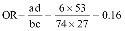
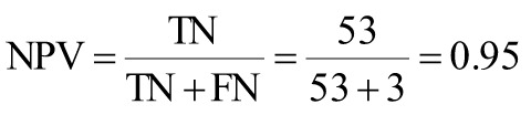
The aim of this article is to determine the efficacy of otoendoscopy during microscopic cholesteatoma surgery on residual cholesteatoma rates postoperatively. The medical records of patients (aged 4-90) with primary acquired cholesteatoma who underwent microscopic cholesteatoma surgery (exclusively transcanal approach or canal wall-up tympano-mastoidectomy) with subsequent otoendoscopic examination (80 ears) for intraoperative cholesteatoma residues were retrospectively reviewed. All cases with mixed microscopic/endoscopic, fully endoscopic, or fully microscopic dissection were excluded, as well as cases where a canal wall-down technique was used. After microscopic cholesteatoma removal, the otoendoscope was used to inspect the middle ear recesses for intraoperative cholesteatoma residues. The intra- and postoperative cholesteatoma residue rate were evaluated. On endoscopic examination, intraoperative cholesteatoma residues were encountered in 24 patients (30%). A total of 30 foci were detected. Most of them were found in the superior retrotympanum (15 foci). In 9 cases an antral remnant guided the surgeon to convert to a canal wall up tympanomastoidectomy. During the postoperative follow-up period, residual cholesteatoma was detected on postoperative magnetic resonance imaging in 6 patients (7.5%). Adding an otoendoscopic examination to microscopic cholesteatoma surgery reduced the postoperative cholesteatoma residues rate (odds ratio=0.16). A negative otoendoscopic examination led to a cholesteatoma residue-free follow-up period in 95% of cases(NPV=0.95). Otoendoscopy is effective in identifying intraoperative cholesteatoma residues after microscopic cholesteatoma surgery. It reduces the postoperative cholesteatoma residue rate, and a negative otoendoscopic examination increases the likelihood of a cholesteatoma residue-free follow-up.


 分享
分享

 求助内容:
求助内容: 应助结果提醒方式:
 扫码关注我们
