Rahman Ud Din, Tahira Nishtar, Xiaoguang Cheng, Haisheng Yang
{"title":"基于磁共振成像模型的 S1 椎体评分是绝经后妇女脂肪-水样骨质疏松变化的指标:一项试点研究。","authors":"Rahman Ud Din, Tahira Nishtar, Xiaoguang Cheng, Haisheng Yang","doi":"10.31616/asj.2024.0116","DOIUrl":null,"url":null,"abstract":"<p><strong>Study design: </strong>A prospective study.</p><p><strong>Purpose: </strong>To assess fat-water-like tissue changes on the 1st sacral vertebra using novel magnetic resonance imaging (MRI) phantombased F- and W-scores and evaluate their diagnostic performances in osteoporosis detection.</p><p><strong>Overview of literature: </strong>Using an uncommonly advanced MRI technique, previous studies have found that fat-water changes were consistent with osteoporosis. The role of routine MRI sequences can be extended in this regard. The S1 vertebra is considered a crucial anatomical site in spine surgeries because it seldom suffers from fractures. Thus, S1 could indicate osteoporotic fat-water changes.</p><p><strong>Methods: </strong>Forty-two female volunteers (aged 62.3±6.3 years) underwent spine examination with both MRI (including a phantom) and dual-energy X-ray absorptiometry (DXA) following ethical approval. MRI phantom-based F- and W-scoreS1 were defined by normalizing S1 vertebral signal intensities (SIs) by coconut oil and water SIs of the phantom on T1- and T2-weighted imaging, respectively. Using receiver operating characteristic analysis, the diagnostic performances of the new scores for evaluating osteoporosis and vertebral fractures were investigated against standard areal bone mineral density measured with DXA (DXA-aBMD).</p><p><strong>Results: </strong>The F-scoreS1 and W-scoreS1 were greater (4.11 and 2.43, respectively) in patients with osteoporosis than those without osteoporosis (3.25 and 1.92, respectively) and achieved areas under the curve (AUCs) of 0.82 and 0.76 (p<0.05), respectively, for osteoporosis detection. Similarly, the mean F-scoreS1 and W-scoreS1 were higher (4.11 and 2.63, respectively) in patients with vertebral fractures than in those without fractures (3.30 and 1.82, respectively) and had greater AUCs (0.90 for W-scoreS1 and 0.74 for F-scoreS1) than DXA-aBMD (AUC, 0.26; p<0.03). In addition, the F- and W-scoreS1 demonstrated a strong correlation (r=0.65, p<0.001).</p><p><strong>Conclusions: </strong>The new S1 vertebral-based MRI scores were developed to detect osteoporotic changes and demonstrated improvements over DXA-aBMD in differentiating patients with vertebral fractures.</p>","PeriodicalId":8555,"journal":{"name":"Asian Spine Journal","volume":" ","pages":"560-569"},"PeriodicalIF":2.7000,"publicationDate":"2024-08-01","publicationTypes":"Journal Article","fieldsOfStudy":null,"isOpenAccess":false,"openAccessPdf":"https://www.ncbi.nlm.nih.gov/pmc/articles/PMC11366554/pdf/","citationCount":"0","resultStr":"{\"title\":\"Magnetic resonance imaging phantom-based S1 vertebral scores are indicators of fat-water-like osteoporotic changes in postmenopausal women: a pilot study.\",\"authors\":\"Rahman Ud Din, Tahira Nishtar, Xiaoguang Cheng, Haisheng Yang\",\"doi\":\"10.31616/asj.2024.0116\",\"DOIUrl\":null,\"url\":null,\"abstract\":\"<p><strong>Study design: </strong>A prospective study.</p><p><strong>Purpose: </strong>To assess fat-water-like tissue changes on the 1st sacral vertebra using novel magnetic resonance imaging (MRI) phantombased F- and W-scores and evaluate their diagnostic performances in osteoporosis detection.</p><p><strong>Overview of literature: </strong>Using an uncommonly advanced MRI technique, previous studies have found that fat-water changes were consistent with osteoporosis. The role of routine MRI sequences can be extended in this regard. The S1 vertebra is considered a crucial anatomical site in spine surgeries because it seldom suffers from fractures. Thus, S1 could indicate osteoporotic fat-water changes.</p><p><strong>Methods: </strong>Forty-two female volunteers (aged 62.3±6.3 years) underwent spine examination with both MRI (including a phantom) and dual-energy X-ray absorptiometry (DXA) following ethical approval. MRI phantom-based F- and W-scoreS1 were defined by normalizing S1 vertebral signal intensities (SIs) by coconut oil and water SIs of the phantom on T1- and T2-weighted imaging, respectively. Using receiver operating characteristic analysis, the diagnostic performances of the new scores for evaluating osteoporosis and vertebral fractures were investigated against standard areal bone mineral density measured with DXA (DXA-aBMD).</p><p><strong>Results: </strong>The F-scoreS1 and W-scoreS1 were greater (4.11 and 2.43, respectively) in patients with osteoporosis than those without osteoporosis (3.25 and 1.92, respectively) and achieved areas under the curve (AUCs) of 0.82 and 0.76 (p<0.05), respectively, for osteoporosis detection. Similarly, the mean F-scoreS1 and W-scoreS1 were higher (4.11 and 2.63, respectively) in patients with vertebral fractures than in those without fractures (3.30 and 1.82, respectively) and had greater AUCs (0.90 for W-scoreS1 and 0.74 for F-scoreS1) than DXA-aBMD (AUC, 0.26; p<0.03). In addition, the F- and W-scoreS1 demonstrated a strong correlation (r=0.65, p<0.001).</p><p><strong>Conclusions: </strong>The new S1 vertebral-based MRI scores were developed to detect osteoporotic changes and demonstrated improvements over DXA-aBMD in differentiating patients with vertebral fractures.</p>\",\"PeriodicalId\":8555,\"journal\":{\"name\":\"Asian Spine Journal\",\"volume\":\" \",\"pages\":\"560-569\"},\"PeriodicalIF\":2.7000,\"publicationDate\":\"2024-08-01\",\"publicationTypes\":\"Journal Article\",\"fieldsOfStudy\":null,\"isOpenAccess\":false,\"openAccessPdf\":\"https://www.ncbi.nlm.nih.gov/pmc/articles/PMC11366554/pdf/\",\"citationCount\":\"0\",\"resultStr\":null,\"platform\":\"Semanticscholar\",\"paperid\":null,\"PeriodicalName\":\"Asian Spine Journal\",\"FirstCategoryId\":\"1085\",\"ListUrlMain\":\"https://doi.org/10.31616/asj.2024.0116\",\"RegionNum\":0,\"RegionCategory\":null,\"ArticlePicture\":[],\"TitleCN\":null,\"AbstractTextCN\":null,\"PMCID\":null,\"EPubDate\":\"2024/8/21 0:00:00\",\"PubModel\":\"Epub\",\"JCR\":\"Q2\",\"JCRName\":\"ORTHOPEDICS\",\"Score\":null,\"Total\":0}","platform":"Semanticscholar","paperid":null,"PeriodicalName":"Asian Spine Journal","FirstCategoryId":"1085","ListUrlMain":"https://doi.org/10.31616/asj.2024.0116","RegionNum":0,"RegionCategory":null,"ArticlePicture":[],"TitleCN":null,"AbstractTextCN":null,"PMCID":null,"EPubDate":"2024/8/21 0:00:00","PubModel":"Epub","JCR":"Q2","JCRName":"ORTHOPEDICS","Score":null,"Total":0}
引用次数: 0
摘要
研究设计目的:使用新型磁共振成像(MRI)幻影 F 值和 W 值评估第 1 骶椎的脂肪水样组织变化,并评估其在骨质疏松症检测中的诊断性能:以往的研究发现,脂肪-水变化与骨质疏松症一致。在这方面,常规 MRI 序列的作用可以得到扩展。S1 椎体被认为是脊柱手术中的关键解剖部位,因为它很少发生骨折。因此,S1 可显示骨质疏松性脂肪-水变化:42名女性志愿者(年龄为62.3±6.3岁)在获得伦理批准后接受了核磁共振成像(包括一个模型)和双能X射线吸收测量(DXA)的脊柱检查。在 T1 和 T2 加权成像中,通过将 S1 椎体信号强度(SI)分别与模型的椰子油和水 SI 进行归一化,定义了基于核磁共振成像模型的 F 值和 W 值S1。通过接收器操作特征分析,研究了新评分在评估骨质疏松症和椎体骨折方面与 DXA 测量的标准骨矿密度(DXA-aBMD)的诊断性能:结果:骨质疏松症患者的 F-scoreS1 和 W-scoreS1 分别为 4.11 和 2.43,高于非骨质疏松症患者(分别为 3.25 和 1.92),曲线下面积(AUC)分别为 0.82 和 0.76(pConclusions):新的基于S1椎体的磁共振成像评分是为检测骨质疏松性变化而开发的,在区分椎体骨折患者方面比DXA-aBMD有所改进。
Magnetic resonance imaging phantom-based S1 vertebral scores are indicators of fat-water-like osteoporotic changes in postmenopausal women: a pilot study.
Study design: A prospective study.
Purpose: To assess fat-water-like tissue changes on the 1st sacral vertebra using novel magnetic resonance imaging (MRI) phantombased F- and W-scores and evaluate their diagnostic performances in osteoporosis detection.
Overview of literature: Using an uncommonly advanced MRI technique, previous studies have found that fat-water changes were consistent with osteoporosis. The role of routine MRI sequences can be extended in this regard. The S1 vertebra is considered a crucial anatomical site in spine surgeries because it seldom suffers from fractures. Thus, S1 could indicate osteoporotic fat-water changes.
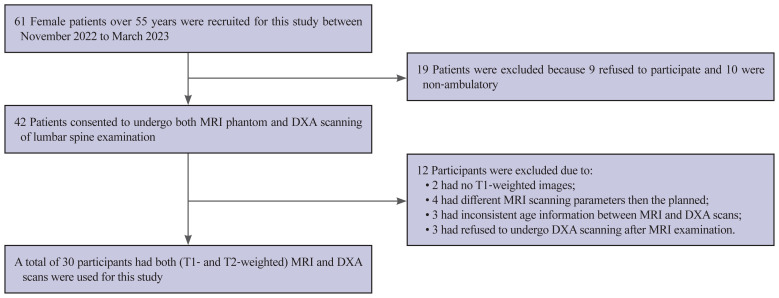
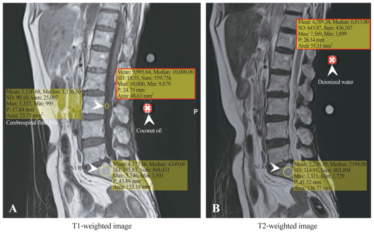
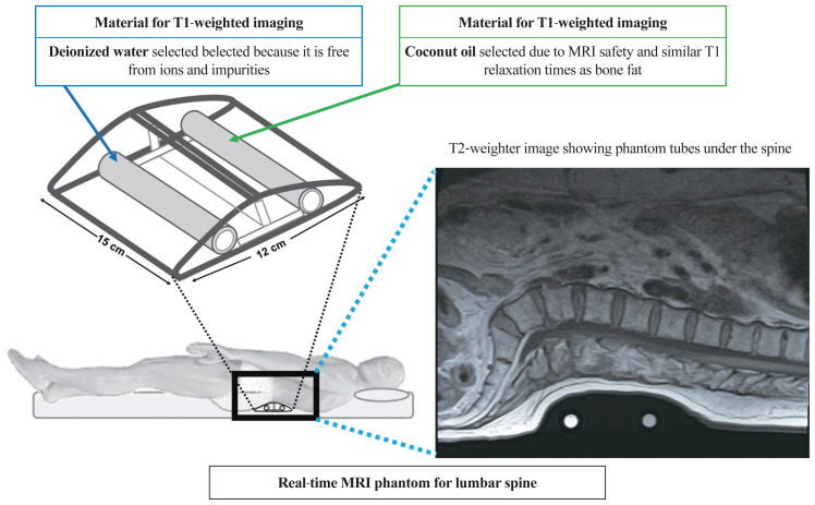
Methods: Forty-two female volunteers (aged 62.3±6.3 years) underwent spine examination with both MRI (including a phantom) and dual-energy X-ray absorptiometry (DXA) following ethical approval. MRI phantom-based F- and W-scoreS1 were defined by normalizing S1 vertebral signal intensities (SIs) by coconut oil and water SIs of the phantom on T1- and T2-weighted imaging, respectively. Using receiver operating characteristic analysis, the diagnostic performances of the new scores for evaluating osteoporosis and vertebral fractures were investigated against standard areal bone mineral density measured with DXA (DXA-aBMD).
Results: The F-scoreS1 and W-scoreS1 were greater (4.11 and 2.43, respectively) in patients with osteoporosis than those without osteoporosis (3.25 and 1.92, respectively) and achieved areas under the curve (AUCs) of 0.82 and 0.76 (p<0.05), respectively, for osteoporosis detection. Similarly, the mean F-scoreS1 and W-scoreS1 were higher (4.11 and 2.63, respectively) in patients with vertebral fractures than in those without fractures (3.30 and 1.82, respectively) and had greater AUCs (0.90 for W-scoreS1 and 0.74 for F-scoreS1) than DXA-aBMD (AUC, 0.26; p<0.03). In addition, the F- and W-scoreS1 demonstrated a strong correlation (r=0.65, p<0.001).
Conclusions: The new S1 vertebral-based MRI scores were developed to detect osteoporotic changes and demonstrated improvements over DXA-aBMD in differentiating patients with vertebral fractures.


 分享
分享
 求助内容:
求助内容: 应助结果提醒方式:
 扫码关注我们
