{"title":"病例报告:一种间充质软骨肉瘤,椎间盘中存在替代性 HEY1::NCOA2 融合。","authors":"Satsuki Kishikawa, Akihide Kondo, Takashi Yao, Tsuyoshi Saito","doi":"10.3389/pore.2024.1611730","DOIUrl":null,"url":null,"abstract":"<p><strong>Introduction: </strong>Mesenchymal chondrosarcoma (MCS) is a rare subtype of chondrosarcoma that occurs at widespread anatomical locations, such as bone, soft tissue, and intracranial sites. The central nervous system (CNS) is one of the most common origins of extraosseous MCS. However, alternative <i>HEY1::NCOA2</i> fusions have not been reported in this tumor.</p><p><strong>Case report: </strong>We report a case of intracranial MCS with <i>HEY1::NCOA2</i> rearrangement. A 52-year-old woman presented with a 15-mm calcified mass around the sella turcica. She initially underwent transsphenoidal surgery for tumor resection and then additional resections for five local recurrences over 5 years. Histologically, the tumor was composed of small round to spindle-shaped cells admixed with well-differentiated hyaline cartilaginous islands. A hemangiopericytoma-like vascular pattern and small sinusoid-like vessels were also observed. RNA sequencing using RNA extracted from formalin-fixed paraffin-embedded samples from the last operation revealed two alternative variants of the <i>HEY1::NCOA2</i> fusion: <i>HEY1</i>(ex4)::<i>NCOA2</i> (ex13) and <i>HEY1</i>(ex4)::<i>NCOA2</i>(ex14). Both variants were confirmed as in-frame fusions using reverse transcription-polymerase chain reaction.</p><p><strong>Discussion: </strong>Cartilaginous components were often not apparent during the recurrences. In addition to the non-typical pathological finding, the correct diagnosis was hampered by the poor RNA quality of the surgical specimens and non-specific STAT6 nuclear staining.</p><p><strong>Conclusion: </strong>This is the first reported case of intracranial MCS with an alternative <i>HEY1::NCOA2</i> fusion.</p>","PeriodicalId":19981,"journal":{"name":"Pathology & Oncology Research","volume":"30 ","pages":"1611730"},"PeriodicalIF":2.3000,"publicationDate":"2024-08-06","publicationTypes":"Journal Article","fieldsOfStudy":null,"isOpenAccess":false,"openAccessPdf":"https://www.ncbi.nlm.nih.gov/pmc/articles/PMC11333213/pdf/","citationCount":"0","resultStr":"{\"title\":\"Case report: A mesenchymal chondrosarcoma with alternative <i>HEY1::NCOA2</i> fusions in the sella turcica.\",\"authors\":\"Satsuki Kishikawa, Akihide Kondo, Takashi Yao, Tsuyoshi Saito\",\"doi\":\"10.3389/pore.2024.1611730\",\"DOIUrl\":null,\"url\":null,\"abstract\":\"<p><strong>Introduction: </strong>Mesenchymal chondrosarcoma (MCS) is a rare subtype of chondrosarcoma that occurs at widespread anatomical locations, such as bone, soft tissue, and intracranial sites. The central nervous system (CNS) is one of the most common origins of extraosseous MCS. However, alternative <i>HEY1::NCOA2</i> fusions have not been reported in this tumor.</p><p><strong>Case report: </strong>We report a case of intracranial MCS with <i>HEY1::NCOA2</i> rearrangement. A 52-year-old woman presented with a 15-mm calcified mass around the sella turcica. She initially underwent transsphenoidal surgery for tumor resection and then additional resections for five local recurrences over 5 years. Histologically, the tumor was composed of small round to spindle-shaped cells admixed with well-differentiated hyaline cartilaginous islands. A hemangiopericytoma-like vascular pattern and small sinusoid-like vessels were also observed. RNA sequencing using RNA extracted from formalin-fixed paraffin-embedded samples from the last operation revealed two alternative variants of the <i>HEY1::NCOA2</i> fusion: <i>HEY1</i>(ex4)::<i>NCOA2</i> (ex13) and <i>HEY1</i>(ex4)::<i>NCOA2</i>(ex14). Both variants were confirmed as in-frame fusions using reverse transcription-polymerase chain reaction.</p><p><strong>Discussion: </strong>Cartilaginous components were often not apparent during the recurrences. In addition to the non-typical pathological finding, the correct diagnosis was hampered by the poor RNA quality of the surgical specimens and non-specific STAT6 nuclear staining.</p><p><strong>Conclusion: </strong>This is the first reported case of intracranial MCS with an alternative <i>HEY1::NCOA2</i> fusion.</p>\",\"PeriodicalId\":19981,\"journal\":{\"name\":\"Pathology & Oncology Research\",\"volume\":\"30 \",\"pages\":\"1611730\"},\"PeriodicalIF\":2.3000,\"publicationDate\":\"2024-08-06\",\"publicationTypes\":\"Journal Article\",\"fieldsOfStudy\":null,\"isOpenAccess\":false,\"openAccessPdf\":\"https://www.ncbi.nlm.nih.gov/pmc/articles/PMC11333213/pdf/\",\"citationCount\":\"0\",\"resultStr\":null,\"platform\":\"Semanticscholar\",\"paperid\":null,\"PeriodicalName\":\"Pathology & Oncology Research\",\"FirstCategoryId\":\"3\",\"ListUrlMain\":\"https://doi.org/10.3389/pore.2024.1611730\",\"RegionNum\":4,\"RegionCategory\":\"医学\",\"ArticlePicture\":[],\"TitleCN\":null,\"AbstractTextCN\":null,\"PMCID\":null,\"EPubDate\":\"2024/1/1 0:00:00\",\"PubModel\":\"eCollection\",\"JCR\":\"Q3\",\"JCRName\":\"ONCOLOGY\",\"Score\":null,\"Total\":0}","platform":"Semanticscholar","paperid":null,"PeriodicalName":"Pathology & Oncology Research","FirstCategoryId":"3","ListUrlMain":"https://doi.org/10.3389/pore.2024.1611730","RegionNum":4,"RegionCategory":"医学","ArticlePicture":[],"TitleCN":null,"AbstractTextCN":null,"PMCID":null,"EPubDate":"2024/1/1 0:00:00","PubModel":"eCollection","JCR":"Q3","JCRName":"ONCOLOGY","Score":null,"Total":0}
Case report: A mesenchymal chondrosarcoma with alternative HEY1::NCOA2 fusions in the sella turcica.
Introduction: Mesenchymal chondrosarcoma (MCS) is a rare subtype of chondrosarcoma that occurs at widespread anatomical locations, such as bone, soft tissue, and intracranial sites. The central nervous system (CNS) is one of the most common origins of extraosseous MCS. However, alternative HEY1::NCOA2 fusions have not been reported in this tumor.
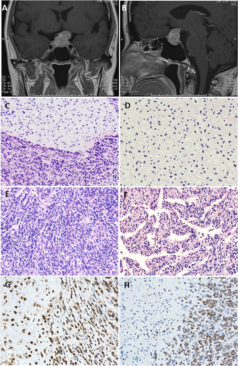
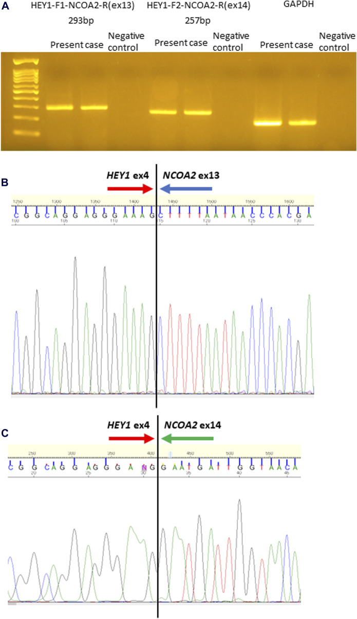
Case report: We report a case of intracranial MCS with HEY1::NCOA2 rearrangement. A 52-year-old woman presented with a 15-mm calcified mass around the sella turcica. She initially underwent transsphenoidal surgery for tumor resection and then additional resections for five local recurrences over 5 years. Histologically, the tumor was composed of small round to spindle-shaped cells admixed with well-differentiated hyaline cartilaginous islands. A hemangiopericytoma-like vascular pattern and small sinusoid-like vessels were also observed. RNA sequencing using RNA extracted from formalin-fixed paraffin-embedded samples from the last operation revealed two alternative variants of the HEY1::NCOA2 fusion: HEY1(ex4)::NCOA2 (ex13) and HEY1(ex4)::NCOA2(ex14). Both variants were confirmed as in-frame fusions using reverse transcription-polymerase chain reaction.
Discussion: Cartilaginous components were often not apparent during the recurrences. In addition to the non-typical pathological finding, the correct diagnosis was hampered by the poor RNA quality of the surgical specimens and non-specific STAT6 nuclear staining.
Conclusion: This is the first reported case of intracranial MCS with an alternative HEY1::NCOA2 fusion.
期刊介绍:
Pathology & Oncology Research (POR) is an interdisciplinary Journal at the interface of pathology and oncology including the preclinical and translational research, diagnostics and therapy. Furthermore, POR is an international forum for the rapid communication of reviews, original research, critical and topical reports with excellence and novelty. Published quarterly, POR is dedicated to keeping scientists informed of developments on the selected biomedical fields bridging the gap between basic research and clinical medicine. It is a special aim for POR to promote pathological and oncological publishing activity of colleagues in the Central and East European region. The journal will be of interest to pathologists, and a broad range of experimental and clinical oncologists, and related experts. POR is supported by an acknowledged international advisory board and the Arányi Fundation for modern pathology.


 分享
分享
 求助内容:
求助内容: 应助结果提醒方式:
 扫码关注我们
