{"title":"人工智能辅助体积各向同性同步交错亮血和黑血检查脑转移瘤。","authors":"Kazufumi Kikuchi, Osamu Togao, Yoshitomo Kikuchi, Koji Yamashita, Daichi Momosaka, Kazunori Fukasawa, Shunsuke Nishimura, Hiroyuki Toyoda, Makoto Obara, Akio Hiwatashi, Kousei Ishigami","doi":"10.1007/s00234-024-03454-4","DOIUrl":null,"url":null,"abstract":"<p><strong>Purpose: </strong>To verify the effectiveness of artificial intelligence-assisted volume isotropic simultaneous interleaved bright-/black-blood examination (AI-VISIBLE) for detecting brain metastases.</p><p><strong>Methods: </strong>This retrospective study was approved by our institutional review board and the requirement for written informed consent was waived. Forty patients were included: 20 patients with and without brain metastases each. Seven independent observers (three radiology residents and four neuroradiologists) participated in two reading sessions: in the first, brain metastases were detected using VISIBLE only; in the second, the results of the first session were comprehensively evaluated by adding AI-VISIBLE information. Sensitivity, diagnostic performance, and false positives/case were evaluated. Diagnostic performance was assessed using a figure-of-merit (FOM). Sensitivity and false positives/case were evaluated using McNemar and paired t-tests, respectively.</p><p><strong>Results: </strong>The McNemar test revealed a significant difference between VISIBLE with/without AI information (P < 0.0001). Significantly higher sensitivity (94.9 ± 1.7% vs. 88.3 ± 5.1%, P = 0.0028) and FOM (0.983 ± 0.009 vs. 0.972 ± 0.013, P = 0.0063) were achieved using VISIBLE with AI information vs. without. No significant difference was observed in false positives/case with and without AI information (0.23 ± 0.19 vs. 0.18 ± 0.15, P = 0.250). AI-assisted results of radiology residents became comparable to results of neuroradiologists (sensitivity, FOM: 85.9 ± 3.4% vs. 90.0 ± 5.9%, 0.969 ± 0.016 vs. 0.974 ± 0.012 without AI information; 94.8 ± 1.3% vs. 95.0 ± 2.1%, 0.977 ± 0.010 vs. 0.988 ± 0.005 with AI information, respectively).</p><p><strong>Conclusion: </strong>AI-VISIBLE improved the sensitivity and performance for diagnosing brain metastases.</p>","PeriodicalId":19422,"journal":{"name":"Neuroradiology","volume":" ","pages":"351-359"},"PeriodicalIF":2.6000,"publicationDate":"2025-02-01","publicationTypes":"Journal Article","fieldsOfStudy":null,"isOpenAccess":false,"openAccessPdf":"https://www.ncbi.nlm.nih.gov/pmc/articles/PMC11893687/pdf/","citationCount":"0","resultStr":"{\"title\":\"Artificial intelligence-assisted volume isotropic simultaneous interleaved bright- and black-blood examination for brain metastases.\",\"authors\":\"Kazufumi Kikuchi, Osamu Togao, Yoshitomo Kikuchi, Koji Yamashita, Daichi Momosaka, Kazunori Fukasawa, Shunsuke Nishimura, Hiroyuki Toyoda, Makoto Obara, Akio Hiwatashi, Kousei Ishigami\",\"doi\":\"10.1007/s00234-024-03454-4\",\"DOIUrl\":null,\"url\":null,\"abstract\":\"<p><strong>Purpose: </strong>To verify the effectiveness of artificial intelligence-assisted volume isotropic simultaneous interleaved bright-/black-blood examination (AI-VISIBLE) for detecting brain metastases.</p><p><strong>Methods: </strong>This retrospective study was approved by our institutional review board and the requirement for written informed consent was waived. Forty patients were included: 20 patients with and without brain metastases each. Seven independent observers (three radiology residents and four neuroradiologists) participated in two reading sessions: in the first, brain metastases were detected using VISIBLE only; in the second, the results of the first session were comprehensively evaluated by adding AI-VISIBLE information. Sensitivity, diagnostic performance, and false positives/case were evaluated. Diagnostic performance was assessed using a figure-of-merit (FOM). Sensitivity and false positives/case were evaluated using McNemar and paired t-tests, respectively.</p><p><strong>Results: </strong>The McNemar test revealed a significant difference between VISIBLE with/without AI information (P < 0.0001). Significantly higher sensitivity (94.9 ± 1.7% vs. 88.3 ± 5.1%, P = 0.0028) and FOM (0.983 ± 0.009 vs. 0.972 ± 0.013, P = 0.0063) were achieved using VISIBLE with AI information vs. without. No significant difference was observed in false positives/case with and without AI information (0.23 ± 0.19 vs. 0.18 ± 0.15, P = 0.250). AI-assisted results of radiology residents became comparable to results of neuroradiologists (sensitivity, FOM: 85.9 ± 3.4% vs. 90.0 ± 5.9%, 0.969 ± 0.016 vs. 0.974 ± 0.012 without AI information; 94.8 ± 1.3% vs. 95.0 ± 2.1%, 0.977 ± 0.010 vs. 0.988 ± 0.005 with AI information, respectively).</p><p><strong>Conclusion: </strong>AI-VISIBLE improved the sensitivity and performance for diagnosing brain metastases.</p>\",\"PeriodicalId\":19422,\"journal\":{\"name\":\"Neuroradiology\",\"volume\":\" \",\"pages\":\"351-359\"},\"PeriodicalIF\":2.6000,\"publicationDate\":\"2025-02-01\",\"publicationTypes\":\"Journal Article\",\"fieldsOfStudy\":null,\"isOpenAccess\":false,\"openAccessPdf\":\"https://www.ncbi.nlm.nih.gov/pmc/articles/PMC11893687/pdf/\",\"citationCount\":\"0\",\"resultStr\":null,\"platform\":\"Semanticscholar\",\"paperid\":null,\"PeriodicalName\":\"Neuroradiology\",\"FirstCategoryId\":\"3\",\"ListUrlMain\":\"https://doi.org/10.1007/s00234-024-03454-4\",\"RegionNum\":3,\"RegionCategory\":\"医学\",\"ArticlePicture\":[],\"TitleCN\":null,\"AbstractTextCN\":null,\"PMCID\":null,\"EPubDate\":\"2024/8/22 0:00:00\",\"PubModel\":\"Epub\",\"JCR\":\"Q2\",\"JCRName\":\"CLINICAL NEUROLOGY\",\"Score\":null,\"Total\":0}","platform":"Semanticscholar","paperid":null,"PeriodicalName":"Neuroradiology","FirstCategoryId":"3","ListUrlMain":"https://doi.org/10.1007/s00234-024-03454-4","RegionNum":3,"RegionCategory":"医学","ArticlePicture":[],"TitleCN":null,"AbstractTextCN":null,"PMCID":null,"EPubDate":"2024/8/22 0:00:00","PubModel":"Epub","JCR":"Q2","JCRName":"CLINICAL NEUROLOGY","Score":null,"Total":0}
Artificial intelligence-assisted volume isotropic simultaneous interleaved bright- and black-blood examination for brain metastases.
Purpose: To verify the effectiveness of artificial intelligence-assisted volume isotropic simultaneous interleaved bright-/black-blood examination (AI-VISIBLE) for detecting brain metastases.
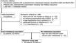
Methods: This retrospective study was approved by our institutional review board and the requirement for written informed consent was waived. Forty patients were included: 20 patients with and without brain metastases each. Seven independent observers (three radiology residents and four neuroradiologists) participated in two reading sessions: in the first, brain metastases were detected using VISIBLE only; in the second, the results of the first session were comprehensively evaluated by adding AI-VISIBLE information. Sensitivity, diagnostic performance, and false positives/case were evaluated. Diagnostic performance was assessed using a figure-of-merit (FOM). Sensitivity and false positives/case were evaluated using McNemar and paired t-tests, respectively.
Results: The McNemar test revealed a significant difference between VISIBLE with/without AI information (P < 0.0001). Significantly higher sensitivity (94.9 ± 1.7% vs. 88.3 ± 5.1%, P = 0.0028) and FOM (0.983 ± 0.009 vs. 0.972 ± 0.013, P = 0.0063) were achieved using VISIBLE with AI information vs. without. No significant difference was observed in false positives/case with and without AI information (0.23 ± 0.19 vs. 0.18 ± 0.15, P = 0.250). AI-assisted results of radiology residents became comparable to results of neuroradiologists (sensitivity, FOM: 85.9 ± 3.4% vs. 90.0 ± 5.9%, 0.969 ± 0.016 vs. 0.974 ± 0.012 without AI information; 94.8 ± 1.3% vs. 95.0 ± 2.1%, 0.977 ± 0.010 vs. 0.988 ± 0.005 with AI information, respectively).
Conclusion: AI-VISIBLE improved the sensitivity and performance for diagnosing brain metastases.
期刊介绍:
Neuroradiology aims to provide state-of-the-art medical and scientific information in the fields of Neuroradiology, Neurosciences, Neurology, Psychiatry, Neurosurgery, and related medical specialities. Neuroradiology as the official Journal of the European Society of Neuroradiology receives submissions from all parts of the world and publishes peer-reviewed original research, comprehensive reviews, educational papers, opinion papers, and short reports on exceptional clinical observations and new technical developments in the field of Neuroimaging and Neurointervention. The journal has subsections for Diagnostic and Interventional Neuroradiology, Advanced Neuroimaging, Paediatric Neuroradiology, Head-Neck-ENT Radiology, Spine Neuroradiology, and for submissions from Japan. Neuroradiology aims to provide new knowledge about and insights into the function and pathology of the human nervous system that may help to better diagnose and treat nervous system diseases. Neuroradiology is a member of the Committee on Publication Ethics (COPE) and follows the COPE core practices. Neuroradiology prefers articles that are free of bias, self-critical regarding limitations, transparent and clear in describing study participants, methods, and statistics, and short in presenting results. Before peer-review all submissions are automatically checked by iThenticate to assess for potential overlap in prior publication.


 分享
分享
 求助内容:
求助内容: 应助结果提醒方式:
 扫码关注我们
