Chang Sun , Yazdan Salimi , Neroladaki Angeliki , Sana Boudabbous , Habib Zaidi
{"title":"用于稀疏视图 CT 重建的高效双域深度学习网络","authors":"Chang Sun , Yazdan Salimi , Neroladaki Angeliki , Sana Boudabbous , Habib Zaidi","doi":"10.1016/j.cmpb.2024.108376","DOIUrl":null,"url":null,"abstract":"<div><h3>Background and Objective</h3><p>We develop an efficient deep-learning based dual-domain reconstruction method for sparse-view CT reconstruction with small training parameters and comparable running time. We aim to investigate the model's capability and its clinical value by performing objective and subjective quality assessments using clinical CT projection data acquired on commercial scanners.</p></div><div><h3>Methods</h3><p>We designed two lightweight networks, namely Sino-Net and Img-Net, to restore the projection and image signal from the DD-Net reconstructed images in the projection and image domains, respectively. The proposed network has small training parameters and comparable running time among dual-domain based reconstruction networks and is easy to train (end-to-end). We prospectively collected clinical thoraco-abdominal CT projection data acquired on a Siemens Biograph 128 Edge CT scanner to train and validate the proposed network. Further, we quantitatively evaluated the CT Hounsfield unit (HU) values on 21 organs and anatomic structures, such as the liver, aorta, and ribcage. We also analyzed the noise properties and compared the signal-to-noise ratio (SNR) and the contrast-to-noise ratio (CNR) of the reconstructed images. Besides, two radiologists conducted the subjective qualitative evaluation including the confidence and conspicuity of anatomic structures, and the overall image quality using a 1–5 likert scoring system.</p></div><div><h3>Results</h3><p>Objective and subjective evaluation showed that the proposed algorithm achieves competitive results in eliminating noise and artifacts, restoring fine structure details, and recovering edges and contours of anatomic structures using 384 views (1/6 sparse rate). The proposed method exhibited good computational cost performance on clinical projection data.</p></div><div><h3>Conclusion</h3><p>This work presents an efficient dual-domain learning network for sparse-view CT reconstruction on raw projection data from a commercial scanner. The study also provides insights for designing an organ-based image quality assessment pipeline for sparse-view reconstruction tasks, potentially benefiting organ-specific dose reduction by sparse-view imaging.</p></div>","PeriodicalId":10624,"journal":{"name":"Computer methods and programs in biomedicine","volume":"256 ","pages":"Article 108376"},"PeriodicalIF":4.8000,"publicationDate":"2024-11-01","publicationTypes":"Journal Article","fieldsOfStudy":null,"isOpenAccess":false,"openAccessPdf":"https://www.sciencedirect.com/science/article/pii/S0169260724003699/pdfft?md5=bb13bc4e96816cac1e0ceb8e8d125a9c&pid=1-s2.0-S0169260724003699-main.pdf","citationCount":"0","resultStr":"{\"title\":\"An efficient dual-domain deep learning network for sparse-view CT reconstruction\",\"authors\":\"Chang Sun , Yazdan Salimi , Neroladaki Angeliki , Sana Boudabbous , Habib Zaidi\",\"doi\":\"10.1016/j.cmpb.2024.108376\",\"DOIUrl\":null,\"url\":null,\"abstract\":\"<div><h3>Background and Objective</h3><p>We develop an efficient deep-learning based dual-domain reconstruction method for sparse-view CT reconstruction with small training parameters and comparable running time. We aim to investigate the model's capability and its clinical value by performing objective and subjective quality assessments using clinical CT projection data acquired on commercial scanners.</p></div><div><h3>Methods</h3><p>We designed two lightweight networks, namely Sino-Net and Img-Net, to restore the projection and image signal from the DD-Net reconstructed images in the projection and image domains, respectively. The proposed network has small training parameters and comparable running time among dual-domain based reconstruction networks and is easy to train (end-to-end). We prospectively collected clinical thoraco-abdominal CT projection data acquired on a Siemens Biograph 128 Edge CT scanner to train and validate the proposed network. Further, we quantitatively evaluated the CT Hounsfield unit (HU) values on 21 organs and anatomic structures, such as the liver, aorta, and ribcage. We also analyzed the noise properties and compared the signal-to-noise ratio (SNR) and the contrast-to-noise ratio (CNR) of the reconstructed images. Besides, two radiologists conducted the subjective qualitative evaluation including the confidence and conspicuity of anatomic structures, and the overall image quality using a 1–5 likert scoring system.</p></div><div><h3>Results</h3><p>Objective and subjective evaluation showed that the proposed algorithm achieves competitive results in eliminating noise and artifacts, restoring fine structure details, and recovering edges and contours of anatomic structures using 384 views (1/6 sparse rate). The proposed method exhibited good computational cost performance on clinical projection data.</p></div><div><h3>Conclusion</h3><p>This work presents an efficient dual-domain learning network for sparse-view CT reconstruction on raw projection data from a commercial scanner. The study also provides insights for designing an organ-based image quality assessment pipeline for sparse-view reconstruction tasks, potentially benefiting organ-specific dose reduction by sparse-view imaging.</p></div>\",\"PeriodicalId\":10624,\"journal\":{\"name\":\"Computer methods and programs in biomedicine\",\"volume\":\"256 \",\"pages\":\"Article 108376\"},\"PeriodicalIF\":4.8000,\"publicationDate\":\"2024-11-01\",\"publicationTypes\":\"Journal Article\",\"fieldsOfStudy\":null,\"isOpenAccess\":false,\"openAccessPdf\":\"https://www.sciencedirect.com/science/article/pii/S0169260724003699/pdfft?md5=bb13bc4e96816cac1e0ceb8e8d125a9c&pid=1-s2.0-S0169260724003699-main.pdf\",\"citationCount\":\"0\",\"resultStr\":null,\"platform\":\"Semanticscholar\",\"paperid\":null,\"PeriodicalName\":\"Computer methods and programs in biomedicine\",\"FirstCategoryId\":\"5\",\"ListUrlMain\":\"https://www.sciencedirect.com/science/article/pii/S0169260724003699\",\"RegionNum\":2,\"RegionCategory\":\"医学\",\"ArticlePicture\":[],\"TitleCN\":null,\"AbstractTextCN\":null,\"PMCID\":null,\"EPubDate\":\"2024/8/16 0:00:00\",\"PubModel\":\"Epub\",\"JCR\":\"Q1\",\"JCRName\":\"COMPUTER SCIENCE, INTERDISCIPLINARY APPLICATIONS\",\"Score\":null,\"Total\":0}","platform":"Semanticscholar","paperid":null,"PeriodicalName":"Computer methods and programs in biomedicine","FirstCategoryId":"5","ListUrlMain":"https://www.sciencedirect.com/science/article/pii/S0169260724003699","RegionNum":2,"RegionCategory":"医学","ArticlePicture":[],"TitleCN":null,"AbstractTextCN":null,"PMCID":null,"EPubDate":"2024/8/16 0:00:00","PubModel":"Epub","JCR":"Q1","JCRName":"COMPUTER SCIENCE, INTERDISCIPLINARY APPLICATIONS","Score":null,"Total":0}
An efficient dual-domain deep learning network for sparse-view CT reconstruction
Background and Objective
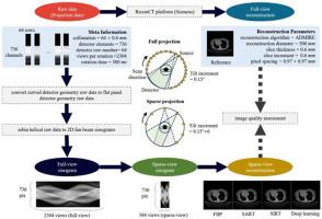
We develop an efficient deep-learning based dual-domain reconstruction method for sparse-view CT reconstruction with small training parameters and comparable running time. We aim to investigate the model's capability and its clinical value by performing objective and subjective quality assessments using clinical CT projection data acquired on commercial scanners.
Methods
We designed two lightweight networks, namely Sino-Net and Img-Net, to restore the projection and image signal from the DD-Net reconstructed images in the projection and image domains, respectively. The proposed network has small training parameters and comparable running time among dual-domain based reconstruction networks and is easy to train (end-to-end). We prospectively collected clinical thoraco-abdominal CT projection data acquired on a Siemens Biograph 128 Edge CT scanner to train and validate the proposed network. Further, we quantitatively evaluated the CT Hounsfield unit (HU) values on 21 organs and anatomic structures, such as the liver, aorta, and ribcage. We also analyzed the noise properties and compared the signal-to-noise ratio (SNR) and the contrast-to-noise ratio (CNR) of the reconstructed images. Besides, two radiologists conducted the subjective qualitative evaluation including the confidence and conspicuity of anatomic structures, and the overall image quality using a 1–5 likert scoring system.
Results
Objective and subjective evaluation showed that the proposed algorithm achieves competitive results in eliminating noise and artifacts, restoring fine structure details, and recovering edges and contours of anatomic structures using 384 views (1/6 sparse rate). The proposed method exhibited good computational cost performance on clinical projection data.
Conclusion
This work presents an efficient dual-domain learning network for sparse-view CT reconstruction on raw projection data from a commercial scanner. The study also provides insights for designing an organ-based image quality assessment pipeline for sparse-view reconstruction tasks, potentially benefiting organ-specific dose reduction by sparse-view imaging.
期刊介绍:
To encourage the development of formal computing methods, and their application in biomedical research and medical practice, by illustration of fundamental principles in biomedical informatics research; to stimulate basic research into application software design; to report the state of research of biomedical information processing projects; to report new computer methodologies applied in biomedical areas; the eventual distribution of demonstrable software to avoid duplication of effort; to provide a forum for discussion and improvement of existing software; to optimize contact between national organizations and regional user groups by promoting an international exchange of information on formal methods, standards and software in biomedicine.
Computer Methods and Programs in Biomedicine covers computing methodology and software systems derived from computing science for implementation in all aspects of biomedical research and medical practice. It is designed to serve: biochemists; biologists; geneticists; immunologists; neuroscientists; pharmacologists; toxicologists; clinicians; epidemiologists; psychiatrists; psychologists; cardiologists; chemists; (radio)physicists; computer scientists; programmers and systems analysts; biomedical, clinical, electrical and other engineers; teachers of medical informatics and users of educational software.


 分享
分享
 求助内容:
求助内容: 应助结果提醒方式:
 扫码关注我们
