Julian Bender, Til Kundlacz, Lucas S.P. Rudden, Melissa Frick, Julia Bieber, Matteo T. Degiacomi, Carla Schmidt
{"title":"Ca2+依赖性脂质偏好决定突触表敏-1 C2A和C2B的动态:实验和模拟的启示","authors":"Julian Bender, Til Kundlacz, Lucas S.P. Rudden, Melissa Frick, Julia Bieber, Matteo T. Degiacomi, Carla Schmidt","doi":"10.1016/j.str.2024.07.017","DOIUrl":null,"url":null,"abstract":"<p>Signal transmission between neurons requires exocytosis of neurotransmitters from the lumen of synaptic vesicles into the synaptic cleft. Following an influx of Ca<sup>2+</sup>, this process is facilitated by the Ca<sup>2+</sup> sensor synaptotagmin-1. The underlying mechanisms involve electrostatic and hydrophobic interactions tuning the lipid preferences of the two C2 domains of synaptotagmin-1; however, the details are still controversially discussed. We, therefore, follow a multidisciplinary approach and characterize lipid and membrane binding of the isolated C2A and C2B domains. We first target interactions with individual lipid species, and then study interactions with model membranes of liposomes. Finally, we perform molecular dynamics simulations to unravel differences in membrane binding. We found that both C2 domains, as a response to Ca<sup>2+</sup>, insert into the lipid membrane; however, C2A adopts a more perpendicular orientation while C2B remains parallel. These findings allow us to propose a mechanism for synaptotagmin-1 during membrane fusion.</p>","PeriodicalId":22168,"journal":{"name":"Structure","volume":"144 1","pages":""},"PeriodicalIF":4.3000,"publicationDate":"2024-08-21","publicationTypes":"Journal Article","fieldsOfStudy":null,"isOpenAccess":false,"openAccessPdf":"","citationCount":"0","resultStr":"{\"title\":\"Ca2+-dependent lipid preferences shape synaptotagmin-1 C2A and C2B dynamics: Insights from experiments and simulations\",\"authors\":\"Julian Bender, Til Kundlacz, Lucas S.P. Rudden, Melissa Frick, Julia Bieber, Matteo T. Degiacomi, Carla Schmidt\",\"doi\":\"10.1016/j.str.2024.07.017\",\"DOIUrl\":null,\"url\":null,\"abstract\":\"<p>Signal transmission between neurons requires exocytosis of neurotransmitters from the lumen of synaptic vesicles into the synaptic cleft. Following an influx of Ca<sup>2+</sup>, this process is facilitated by the Ca<sup>2+</sup> sensor synaptotagmin-1. The underlying mechanisms involve electrostatic and hydrophobic interactions tuning the lipid preferences of the two C2 domains of synaptotagmin-1; however, the details are still controversially discussed. We, therefore, follow a multidisciplinary approach and characterize lipid and membrane binding of the isolated C2A and C2B domains. We first target interactions with individual lipid species, and then study interactions with model membranes of liposomes. Finally, we perform molecular dynamics simulations to unravel differences in membrane binding. We found that both C2 domains, as a response to Ca<sup>2+</sup>, insert into the lipid membrane; however, C2A adopts a more perpendicular orientation while C2B remains parallel. These findings allow us to propose a mechanism for synaptotagmin-1 during membrane fusion.</p>\",\"PeriodicalId\":22168,\"journal\":{\"name\":\"Structure\",\"volume\":\"144 1\",\"pages\":\"\"},\"PeriodicalIF\":4.3000,\"publicationDate\":\"2024-08-21\",\"publicationTypes\":\"Journal Article\",\"fieldsOfStudy\":null,\"isOpenAccess\":false,\"openAccessPdf\":\"\",\"citationCount\":\"0\",\"resultStr\":null,\"platform\":\"Semanticscholar\",\"paperid\":null,\"PeriodicalName\":\"Structure\",\"FirstCategoryId\":\"99\",\"ListUrlMain\":\"https://doi.org/10.1016/j.str.2024.07.017\",\"RegionNum\":2,\"RegionCategory\":\"生物学\",\"ArticlePicture\":[],\"TitleCN\":null,\"AbstractTextCN\":null,\"PMCID\":null,\"EPubDate\":\"\",\"PubModel\":\"\",\"JCR\":\"Q2\",\"JCRName\":\"BIOCHEMISTRY & MOLECULAR BIOLOGY\",\"Score\":null,\"Total\":0}","platform":"Semanticscholar","paperid":null,"PeriodicalName":"Structure","FirstCategoryId":"99","ListUrlMain":"https://doi.org/10.1016/j.str.2024.07.017","RegionNum":2,"RegionCategory":"生物学","ArticlePicture":[],"TitleCN":null,"AbstractTextCN":null,"PMCID":null,"EPubDate":"","PubModel":"","JCR":"Q2","JCRName":"BIOCHEMISTRY & MOLECULAR BIOLOGY","Score":null,"Total":0}
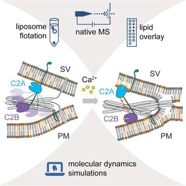
Ca2+-dependent lipid preferences shape synaptotagmin-1 C2A and C2B dynamics: Insights from experiments and simulations
Signal transmission between neurons requires exocytosis of neurotransmitters from the lumen of synaptic vesicles into the synaptic cleft. Following an influx of Ca2+, this process is facilitated by the Ca2+ sensor synaptotagmin-1. The underlying mechanisms involve electrostatic and hydrophobic interactions tuning the lipid preferences of the two C2 domains of synaptotagmin-1; however, the details are still controversially discussed. We, therefore, follow a multidisciplinary approach and characterize lipid and membrane binding of the isolated C2A and C2B domains. We first target interactions with individual lipid species, and then study interactions with model membranes of liposomes. Finally, we perform molecular dynamics simulations to unravel differences in membrane binding. We found that both C2 domains, as a response to Ca2+, insert into the lipid membrane; however, C2A adopts a more perpendicular orientation while C2B remains parallel. These findings allow us to propose a mechanism for synaptotagmin-1 during membrane fusion.
期刊介绍:
Structure aims to publish papers of exceptional interest in the field of structural biology. The journal strives to be essential reading for structural biologists, as well as biologists and biochemists that are interested in macromolecular structure and function. Structure strongly encourages the submission of manuscripts that present structural and molecular insights into biological function and mechanism. Other reports that address fundamental questions in structural biology, such as structure-based examinations of protein evolution, folding, and/or design, will also be considered. We will consider the application of any method, experimental or computational, at high or low resolution, to conduct structural investigations, as long as the method is appropriate for the biological, functional, and mechanistic question(s) being addressed. Likewise, reports describing single-molecule analysis of biological mechanisms are welcome.
In general, the editors encourage submission of experimental structural studies that are enriched by an analysis of structure-activity relationships and will not consider studies that solely report structural information unless the structure or analysis is of exceptional and broad interest. Studies reporting only homology models, de novo models, or molecular dynamics simulations are also discouraged unless the models are informed by or validated by novel experimental data; rationalization of a large body of existing experimental evidence and making testable predictions based on a model or simulation is often not considered sufficient.


 分享
分享
 求助内容:
求助内容: 应助结果提醒方式:
 扫码关注我们
