{"title":"经计算机断层扫描血管造影术诊断,右侧花瓣状颈内动脉(ICA)走向异常,伴有右侧 1 型原跖动脉和双侧枕动脉从 ICA 上发出。","authors":"Akira Uchino, Kazuo Tokushige","doi":"10.1007/s00276-024-03466-y","DOIUrl":null,"url":null,"abstract":"<p><strong>Purpose: </strong>To describe a case of multiple extremely rare cervical arterial variations.</p><p><strong>Methods: </strong>A 55-year-old man with a tentative diagnosis of right internal carotid artery (ICA) stenosis was examined using computed tomography (CT) angiography for the evaluation of vascular lesions in the neck and head region. A 64-slice CT machine was used.</p><p><strong>Results: </strong>On CT angiography, there was laterally located and narrowed petrous segment of the right ICA, indicative of aberrant course of the petrous ICA. Right vertebral artery (VA) was small in caliber and a relatively large anomalous artery arose from the proximal right ICA. This anomalous artery entered the posterior fossa via the foramen magnum, indicative of a type 1 proatlantal artery. Right occipital artery (OA) arose from the proximal ICA. The left OA also arose from the proximal ICA.</p><p><strong>Conclusion: </strong>An aberrant course of the petrous ICA is an extremely rare arterial variation which is formed by segmental agenesis of the cervical ICA, and the collateral channel passes through the middle ear cavity. It can be dangerous during middle ear surgery. The type 1 proatlantal artery is also an extremely rare arterial variation formed by the persistence of the proatlantal intersegmental artery. It is clinically significant because of its unique blood flow from the carotid system to the vertebrobasilar system. The OA rarely arises from the proximal ICA. Identification of these cervical arterial variations before surgery and vascular intervention are important to avoid complications during the procedure.</p>","PeriodicalId":49461,"journal":{"name":"Surgical and Radiologic Anatomy","volume":" ","pages":"1615-1619"},"PeriodicalIF":1.2000,"publicationDate":"2024-10-01","publicationTypes":"Journal Article","fieldsOfStudy":null,"isOpenAccess":false,"openAccessPdf":"","citationCount":"0","resultStr":"{\"title\":\"Aberrant course of the right petrous internal carotid artery (ICA) associated with right type 1 proatlantal artery and bilateral occipital arteries arising from the ICA diagnosed by computed tomography angiography.\",\"authors\":\"Akira Uchino, Kazuo Tokushige\",\"doi\":\"10.1007/s00276-024-03466-y\",\"DOIUrl\":null,\"url\":null,\"abstract\":\"<p><strong>Purpose: </strong>To describe a case of multiple extremely rare cervical arterial variations.</p><p><strong>Methods: </strong>A 55-year-old man with a tentative diagnosis of right internal carotid artery (ICA) stenosis was examined using computed tomography (CT) angiography for the evaluation of vascular lesions in the neck and head region. A 64-slice CT machine was used.</p><p><strong>Results: </strong>On CT angiography, there was laterally located and narrowed petrous segment of the right ICA, indicative of aberrant course of the petrous ICA. Right vertebral artery (VA) was small in caliber and a relatively large anomalous artery arose from the proximal right ICA. This anomalous artery entered the posterior fossa via the foramen magnum, indicative of a type 1 proatlantal artery. Right occipital artery (OA) arose from the proximal ICA. The left OA also arose from the proximal ICA.</p><p><strong>Conclusion: </strong>An aberrant course of the petrous ICA is an extremely rare arterial variation which is formed by segmental agenesis of the cervical ICA, and the collateral channel passes through the middle ear cavity. It can be dangerous during middle ear surgery. The type 1 proatlantal artery is also an extremely rare arterial variation formed by the persistence of the proatlantal intersegmental artery. It is clinically significant because of its unique blood flow from the carotid system to the vertebrobasilar system. The OA rarely arises from the proximal ICA. Identification of these cervical arterial variations before surgery and vascular intervention are important to avoid complications during the procedure.</p>\",\"PeriodicalId\":49461,\"journal\":{\"name\":\"Surgical and Radiologic Anatomy\",\"volume\":\" \",\"pages\":\"1615-1619\"},\"PeriodicalIF\":1.2000,\"publicationDate\":\"2024-10-01\",\"publicationTypes\":\"Journal Article\",\"fieldsOfStudy\":null,\"isOpenAccess\":false,\"openAccessPdf\":\"\",\"citationCount\":\"0\",\"resultStr\":null,\"platform\":\"Semanticscholar\",\"paperid\":null,\"PeriodicalName\":\"Surgical and Radiologic Anatomy\",\"FirstCategoryId\":\"3\",\"ListUrlMain\":\"https://doi.org/10.1007/s00276-024-03466-y\",\"RegionNum\":4,\"RegionCategory\":\"医学\",\"ArticlePicture\":[],\"TitleCN\":null,\"AbstractTextCN\":null,\"PMCID\":null,\"EPubDate\":\"2024/8/23 0:00:00\",\"PubModel\":\"Epub\",\"JCR\":\"Q2\",\"JCRName\":\"Medicine\",\"Score\":null,\"Total\":0}","platform":"Semanticscholar","paperid":null,"PeriodicalName":"Surgical and Radiologic Anatomy","FirstCategoryId":"3","ListUrlMain":"https://doi.org/10.1007/s00276-024-03466-y","RegionNum":4,"RegionCategory":"医学","ArticlePicture":[],"TitleCN":null,"AbstractTextCN":null,"PMCID":null,"EPubDate":"2024/8/23 0:00:00","PubModel":"Epub","JCR":"Q2","JCRName":"Medicine","Score":null,"Total":0}
Aberrant course of the right petrous internal carotid artery (ICA) associated with right type 1 proatlantal artery and bilateral occipital arteries arising from the ICA diagnosed by computed tomography angiography.
Purpose: To describe a case of multiple extremely rare cervical arterial variations.
Methods: A 55-year-old man with a tentative diagnosis of right internal carotid artery (ICA) stenosis was examined using computed tomography (CT) angiography for the evaluation of vascular lesions in the neck and head region. A 64-slice CT machine was used.
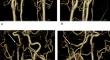
Results: On CT angiography, there was laterally located and narrowed petrous segment of the right ICA, indicative of aberrant course of the petrous ICA. Right vertebral artery (VA) was small in caliber and a relatively large anomalous artery arose from the proximal right ICA. This anomalous artery entered the posterior fossa via the foramen magnum, indicative of a type 1 proatlantal artery. Right occipital artery (OA) arose from the proximal ICA. The left OA also arose from the proximal ICA.
Conclusion: An aberrant course of the petrous ICA is an extremely rare arterial variation which is formed by segmental agenesis of the cervical ICA, and the collateral channel passes through the middle ear cavity. It can be dangerous during middle ear surgery. The type 1 proatlantal artery is also an extremely rare arterial variation formed by the persistence of the proatlantal intersegmental artery. It is clinically significant because of its unique blood flow from the carotid system to the vertebrobasilar system. The OA rarely arises from the proximal ICA. Identification of these cervical arterial variations before surgery and vascular intervention are important to avoid complications during the procedure.
期刊介绍:
Anatomy is a morphological science which cannot fail to interest the clinician. The practical application of anatomical research to clinical problems necessitates special adaptation and selectivity in choosing from numerous international works. Although there is a tendency to believe that meaningful advances in anatomy are unlikely, constant revision is necessary. Surgical and Radiologic Anatomy, the first international journal of Clinical anatomy has been created in this spirit.
Its goal is to serve clinicians, regardless of speciality-physicians, surgeons, radiologists or other specialists-as an indispensable aid with which they can improve their knowledge of anatomy. Each issue includes: Original papers, review articles, articles on the anatomical bases of medical, surgical and radiological techniques, articles of normal radiologic anatomy, brief reviews of anatomical publications of clinical interest.
Particular attention is given to high quality illustrations, which are indispensable for a better understanding of anatomical problems.
Surgical and Radiologic Anatomy is a journal written by anatomists for clinicians with a special interest in anatomy.


 分享
分享
 求助内容:
求助内容: 应助结果提醒方式:
 扫码关注我们
