{"title":"基于多参数双能量非对比 CT 的良性和恶性肝脏病变鉴别预测模型。","authors":"Takashi Ota, Hiromitsu Onishi, Hideyuki Fukui, Takahiro Tsuboyama, Atsushi Nakamoto, Toru Honda, Shohei Matsumoto, Mitsuaki Tatsumi, Noriyuki Tomiyama","doi":"10.1007/s00330-024-11024-8","DOIUrl":null,"url":null,"abstract":"<p><strong>Objectives: </strong>To create prediction models (PMs) for distinguishing between benign and malignant liver lesions using quantitative data from dual-energy CT (DECT) without contrast agents.</p><p><strong>Materials and methods: </strong>This retrospective study included patients with liver lesions who underwent DECT, including non-contrast-enhanced scans. Benign lesions included hepatic hemangioma, whereas malignant lesions included hepatocellular carcinoma, metastatic liver cancer, and intrahepatic cholangiocellular carcinoma. Patients were divided into derivation and validation groups. In the derivation group, two radiologists calculated ten multiparametric data using univariate and multivariate logistic regression to generate PMs. In the validation group, two additional radiologists measured the parameters to assess the diagnostic performance of PMs.</p><p><strong>Results: </strong>The study included 121 consecutive patients (mean age 67.4 ± 13.8 years, 80 males), with 97 in the derivation group (25 benign and 72 malignant) and 24 in the validation group (7 benign and 17 malignant). Oversampling increased the benign lesion sample to 75, equalizing the malignant group for building PMs. All parameters were statistically significant in univariate analysis (all p < 0.05), leading to the creation of five PMs in multivariate analysis. The area under the curve for the five PMs of two observers was as follows: PM1 (slope K, blood) = 0.76, 0.74; PM2 (slope K, fat) = 0.55, 0.51; PM3 (effective-Z difference, blood) = 0.75, 0.72; PM4 (slope K, blood, fat) = 0.82, 0.78; and PM5 (slope K, effective-Z difference, blood) = 0.90, 0.87. PM5 yielded the best diagnostic performance.</p><p><strong>Conclusion: </strong>Multiparametric non-contrast-enhanced DECT is a highly effective method for distinguishing between liver lesions.</p><p><strong>Clinical relevance statement: </strong>The utilization of non-contrast-enhanced DECT is extremely useful for distinguishing between benign and malignant liver lesions. This approach enables physicians to plan better treatment strategies, alleviating concerns associated with contrast allergy, contrast-induced nephropathy, radiation exposure, and excessive medical expenses.</p><p><strong>Key points: </strong>Distinguishing benign from malignant liver lesions with non-contrast-enhanced CT would be desirable. This model, incorporating slope K, effective Z, and blood quantification, distinguished benign from malignant liver lesions. Non-contrast-enhanced DECT has benefits, particularly in patients with an iodine allergy, renal failure, or asthma.</p>","PeriodicalId":12076,"journal":{"name":"European Radiology","volume":" ","pages":"1361-1377"},"PeriodicalIF":4.7000,"publicationDate":"2025-03-01","publicationTypes":"Journal Article","fieldsOfStudy":null,"isOpenAccess":false,"openAccessPdf":"https://www.ncbi.nlm.nih.gov/pmc/articles/PMC11836082/pdf/","citationCount":"0","resultStr":"{\"title\":\"Prediction models for differentiating benign from malignant liver lesions based on multiparametric dual-energy non-contrast CT.\",\"authors\":\"Takashi Ota, Hiromitsu Onishi, Hideyuki Fukui, Takahiro Tsuboyama, Atsushi Nakamoto, Toru Honda, Shohei Matsumoto, Mitsuaki Tatsumi, Noriyuki Tomiyama\",\"doi\":\"10.1007/s00330-024-11024-8\",\"DOIUrl\":null,\"url\":null,\"abstract\":\"<p><strong>Objectives: </strong>To create prediction models (PMs) for distinguishing between benign and malignant liver lesions using quantitative data from dual-energy CT (DECT) without contrast agents.</p><p><strong>Materials and methods: </strong>This retrospective study included patients with liver lesions who underwent DECT, including non-contrast-enhanced scans. Benign lesions included hepatic hemangioma, whereas malignant lesions included hepatocellular carcinoma, metastatic liver cancer, and intrahepatic cholangiocellular carcinoma. Patients were divided into derivation and validation groups. In the derivation group, two radiologists calculated ten multiparametric data using univariate and multivariate logistic regression to generate PMs. In the validation group, two additional radiologists measured the parameters to assess the diagnostic performance of PMs.</p><p><strong>Results: </strong>The study included 121 consecutive patients (mean age 67.4 ± 13.8 years, 80 males), with 97 in the derivation group (25 benign and 72 malignant) and 24 in the validation group (7 benign and 17 malignant). Oversampling increased the benign lesion sample to 75, equalizing the malignant group for building PMs. All parameters were statistically significant in univariate analysis (all p < 0.05), leading to the creation of five PMs in multivariate analysis. The area under the curve for the five PMs of two observers was as follows: PM1 (slope K, blood) = 0.76, 0.74; PM2 (slope K, fat) = 0.55, 0.51; PM3 (effective-Z difference, blood) = 0.75, 0.72; PM4 (slope K, blood, fat) = 0.82, 0.78; and PM5 (slope K, effective-Z difference, blood) = 0.90, 0.87. PM5 yielded the best diagnostic performance.</p><p><strong>Conclusion: </strong>Multiparametric non-contrast-enhanced DECT is a highly effective method for distinguishing between liver lesions.</p><p><strong>Clinical relevance statement: </strong>The utilization of non-contrast-enhanced DECT is extremely useful for distinguishing between benign and malignant liver lesions. This approach enables physicians to plan better treatment strategies, alleviating concerns associated with contrast allergy, contrast-induced nephropathy, radiation exposure, and excessive medical expenses.</p><p><strong>Key points: </strong>Distinguishing benign from malignant liver lesions with non-contrast-enhanced CT would be desirable. This model, incorporating slope K, effective Z, and blood quantification, distinguished benign from malignant liver lesions. Non-contrast-enhanced DECT has benefits, particularly in patients with an iodine allergy, renal failure, or asthma.</p>\",\"PeriodicalId\":12076,\"journal\":{\"name\":\"European Radiology\",\"volume\":\" \",\"pages\":\"1361-1377\"},\"PeriodicalIF\":4.7000,\"publicationDate\":\"2025-03-01\",\"publicationTypes\":\"Journal Article\",\"fieldsOfStudy\":null,\"isOpenAccess\":false,\"openAccessPdf\":\"https://www.ncbi.nlm.nih.gov/pmc/articles/PMC11836082/pdf/\",\"citationCount\":\"0\",\"resultStr\":null,\"platform\":\"Semanticscholar\",\"paperid\":null,\"PeriodicalName\":\"European Radiology\",\"FirstCategoryId\":\"3\",\"ListUrlMain\":\"https://doi.org/10.1007/s00330-024-11024-8\",\"RegionNum\":2,\"RegionCategory\":\"医学\",\"ArticlePicture\":[],\"TitleCN\":null,\"AbstractTextCN\":null,\"PMCID\":null,\"EPubDate\":\"2024/8/26 0:00:00\",\"PubModel\":\"Epub\",\"JCR\":\"Q1\",\"JCRName\":\"RADIOLOGY, NUCLEAR MEDICINE & MEDICAL IMAGING\",\"Score\":null,\"Total\":0}","platform":"Semanticscholar","paperid":null,"PeriodicalName":"European Radiology","FirstCategoryId":"3","ListUrlMain":"https://doi.org/10.1007/s00330-024-11024-8","RegionNum":2,"RegionCategory":"医学","ArticlePicture":[],"TitleCN":null,"AbstractTextCN":null,"PMCID":null,"EPubDate":"2024/8/26 0:00:00","PubModel":"Epub","JCR":"Q1","JCRName":"RADIOLOGY, NUCLEAR MEDICINE & MEDICAL IMAGING","Score":null,"Total":0}
Prediction models for differentiating benign from malignant liver lesions based on multiparametric dual-energy non-contrast CT.
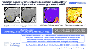
Objectives: To create prediction models (PMs) for distinguishing between benign and malignant liver lesions using quantitative data from dual-energy CT (DECT) without contrast agents.
Materials and methods: This retrospective study included patients with liver lesions who underwent DECT, including non-contrast-enhanced scans. Benign lesions included hepatic hemangioma, whereas malignant lesions included hepatocellular carcinoma, metastatic liver cancer, and intrahepatic cholangiocellular carcinoma. Patients were divided into derivation and validation groups. In the derivation group, two radiologists calculated ten multiparametric data using univariate and multivariate logistic regression to generate PMs. In the validation group, two additional radiologists measured the parameters to assess the diagnostic performance of PMs.
Results: The study included 121 consecutive patients (mean age 67.4 ± 13.8 years, 80 males), with 97 in the derivation group (25 benign and 72 malignant) and 24 in the validation group (7 benign and 17 malignant). Oversampling increased the benign lesion sample to 75, equalizing the malignant group for building PMs. All parameters were statistically significant in univariate analysis (all p < 0.05), leading to the creation of five PMs in multivariate analysis. The area under the curve for the five PMs of two observers was as follows: PM1 (slope K, blood) = 0.76, 0.74; PM2 (slope K, fat) = 0.55, 0.51; PM3 (effective-Z difference, blood) = 0.75, 0.72; PM4 (slope K, blood, fat) = 0.82, 0.78; and PM5 (slope K, effective-Z difference, blood) = 0.90, 0.87. PM5 yielded the best diagnostic performance.
Conclusion: Multiparametric non-contrast-enhanced DECT is a highly effective method for distinguishing between liver lesions.
Clinical relevance statement: The utilization of non-contrast-enhanced DECT is extremely useful for distinguishing between benign and malignant liver lesions. This approach enables physicians to plan better treatment strategies, alleviating concerns associated with contrast allergy, contrast-induced nephropathy, radiation exposure, and excessive medical expenses.
Key points: Distinguishing benign from malignant liver lesions with non-contrast-enhanced CT would be desirable. This model, incorporating slope K, effective Z, and blood quantification, distinguished benign from malignant liver lesions. Non-contrast-enhanced DECT has benefits, particularly in patients with an iodine allergy, renal failure, or asthma.
期刊介绍:
European Radiology (ER) continuously updates scientific knowledge in radiology by publication of strong original articles and state-of-the-art reviews written by leading radiologists. A well balanced combination of review articles, original papers, short communications from European radiological congresses and information on society matters makes ER an indispensable source for current information in this field.
This is the Journal of the European Society of Radiology, and the official journal of a number of societies.
From 2004-2008 supplements to European Radiology were published under its companion, European Radiology Supplements, ISSN 1613-3749.


 分享
分享
 求助内容:
求助内容: 应助结果提醒方式:
 扫码关注我们
