{"title":"磁共振成像头部运动在整个发育过程中的变化:典型发育和多动症。","authors":"Phoebe Thomson, Vanessa Loosley, Emily Friedel, Timothy J Silk","doi":"10.1007/s11682-024-00910-w","DOIUrl":null,"url":null,"abstract":"<p><p>Head motion is a major confounding variable for magnetic resonance imaging (MRI) analysis, and is commonly seen in individuals with neurodevelopmental disorders such as attention deficit hyperactivity disorder (ADHD). This study investigated the trajectory of change in head motion in typically developing children and children with ADHD, and examined possible altered trajectories in head motion between children with remitted and persistent ADHD. 105 children with ADHD and 84 controls completed diffusion and resting-state functional MRI scans at up to three waves over ages 9-14 years. In-scanner head motion was calculated using framewise displacement, and longitudinal trajectories analyzed using generalized additive mixed modelling. Results revealed a significant age effect on framewise displacement where head motion decreased as age increased during both diffusion (p < .001) and resting-state functional MRI (p < .001). A significant effect of group was also observed; children with ADHD displayed greater framewise displacement than controls over the age range (diffusion MRI p = .036, functional MRI p = .004). Further analyses revealed continued elevation in head motion in children in remission from ADHD (diffusion MRI p = .020, functional MRI p = .011) compared to controls. Rates of change in head motion did not significantly differ between diagnostic groups. Findings indicate a critical link between in-scanner head motion and developmental age within children regardless of ADHD diagnosis, important to consider in studies of neurodevelopment. Findings also suggest change in head motion with age does not differ between individuals with remitted and persistent ADHD, adding further evidence that behavioral manifestations of ADHD may continue despite clinical remission.</p>","PeriodicalId":9192,"journal":{"name":"Brain Imaging and Behavior","volume":" ","pages":"1144-1152"},"PeriodicalIF":2.4000,"publicationDate":"2024-10-01","publicationTypes":"Journal Article","fieldsOfStudy":null,"isOpenAccess":false,"openAccessPdf":"https://www.ncbi.nlm.nih.gov/pmc/articles/PMC11582210/pdf/","citationCount":"0","resultStr":"{\"title\":\"Changes in MRI head motion across development: typical development and ADHD.\",\"authors\":\"Phoebe Thomson, Vanessa Loosley, Emily Friedel, Timothy J Silk\",\"doi\":\"10.1007/s11682-024-00910-w\",\"DOIUrl\":null,\"url\":null,\"abstract\":\"<p><p>Head motion is a major confounding variable for magnetic resonance imaging (MRI) analysis, and is commonly seen in individuals with neurodevelopmental disorders such as attention deficit hyperactivity disorder (ADHD). This study investigated the trajectory of change in head motion in typically developing children and children with ADHD, and examined possible altered trajectories in head motion between children with remitted and persistent ADHD. 105 children with ADHD and 84 controls completed diffusion and resting-state functional MRI scans at up to three waves over ages 9-14 years. In-scanner head motion was calculated using framewise displacement, and longitudinal trajectories analyzed using generalized additive mixed modelling. Results revealed a significant age effect on framewise displacement where head motion decreased as age increased during both diffusion (p < .001) and resting-state functional MRI (p < .001). A significant effect of group was also observed; children with ADHD displayed greater framewise displacement than controls over the age range (diffusion MRI p = .036, functional MRI p = .004). Further analyses revealed continued elevation in head motion in children in remission from ADHD (diffusion MRI p = .020, functional MRI p = .011) compared to controls. Rates of change in head motion did not significantly differ between diagnostic groups. Findings indicate a critical link between in-scanner head motion and developmental age within children regardless of ADHD diagnosis, important to consider in studies of neurodevelopment. Findings also suggest change in head motion with age does not differ between individuals with remitted and persistent ADHD, adding further evidence that behavioral manifestations of ADHD may continue despite clinical remission.</p>\",\"PeriodicalId\":9192,\"journal\":{\"name\":\"Brain Imaging and Behavior\",\"volume\":\" \",\"pages\":\"1144-1152\"},\"PeriodicalIF\":2.4000,\"publicationDate\":\"2024-10-01\",\"publicationTypes\":\"Journal Article\",\"fieldsOfStudy\":null,\"isOpenAccess\":false,\"openAccessPdf\":\"https://www.ncbi.nlm.nih.gov/pmc/articles/PMC11582210/pdf/\",\"citationCount\":\"0\",\"resultStr\":null,\"platform\":\"Semanticscholar\",\"paperid\":null,\"PeriodicalName\":\"Brain Imaging and Behavior\",\"FirstCategoryId\":\"3\",\"ListUrlMain\":\"https://doi.org/10.1007/s11682-024-00910-w\",\"RegionNum\":3,\"RegionCategory\":\"医学\",\"ArticlePicture\":[],\"TitleCN\":null,\"AbstractTextCN\":null,\"PMCID\":null,\"EPubDate\":\"2024/8/27 0:00:00\",\"PubModel\":\"Epub\",\"JCR\":\"Q2\",\"JCRName\":\"NEUROIMAGING\",\"Score\":null,\"Total\":0}","platform":"Semanticscholar","paperid":null,"PeriodicalName":"Brain Imaging and Behavior","FirstCategoryId":"3","ListUrlMain":"https://doi.org/10.1007/s11682-024-00910-w","RegionNum":3,"RegionCategory":"医学","ArticlePicture":[],"TitleCN":null,"AbstractTextCN":null,"PMCID":null,"EPubDate":"2024/8/27 0:00:00","PubModel":"Epub","JCR":"Q2","JCRName":"NEUROIMAGING","Score":null,"Total":0}
Changes in MRI head motion across development: typical development and ADHD.
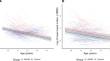
Head motion is a major confounding variable for magnetic resonance imaging (MRI) analysis, and is commonly seen in individuals with neurodevelopmental disorders such as attention deficit hyperactivity disorder (ADHD). This study investigated the trajectory of change in head motion in typically developing children and children with ADHD, and examined possible altered trajectories in head motion between children with remitted and persistent ADHD. 105 children with ADHD and 84 controls completed diffusion and resting-state functional MRI scans at up to three waves over ages 9-14 years. In-scanner head motion was calculated using framewise displacement, and longitudinal trajectories analyzed using generalized additive mixed modelling. Results revealed a significant age effect on framewise displacement where head motion decreased as age increased during both diffusion (p < .001) and resting-state functional MRI (p < .001). A significant effect of group was also observed; children with ADHD displayed greater framewise displacement than controls over the age range (diffusion MRI p = .036, functional MRI p = .004). Further analyses revealed continued elevation in head motion in children in remission from ADHD (diffusion MRI p = .020, functional MRI p = .011) compared to controls. Rates of change in head motion did not significantly differ between diagnostic groups. Findings indicate a critical link between in-scanner head motion and developmental age within children regardless of ADHD diagnosis, important to consider in studies of neurodevelopment. Findings also suggest change in head motion with age does not differ between individuals with remitted and persistent ADHD, adding further evidence that behavioral manifestations of ADHD may continue despite clinical remission.
期刊介绍:
Brain Imaging and Behavior is a bi-monthly, peer-reviewed journal, that publishes clinically relevant research using neuroimaging approaches to enhance our understanding of disorders of higher brain function. The journal is targeted at clinicians and researchers in fields concerned with human brain-behavior relationships, such as neuropsychology, psychiatry, neurology, neurosurgery, rehabilitation, and cognitive neuroscience.


 分享
分享
 求助内容:
求助内容: 应助结果提醒方式:
 扫码关注我们
