{"title":"评估自组装肽和氟化物清漆单独或与激光照射相结合对人工釉质龋齿的影响:SEM/EDS 和显微 CT 研究。","authors":"Aslı Aşık, Özant Önçağ","doi":"10.1007/s00784-024-05901-1","DOIUrl":null,"url":null,"abstract":"<p><strong>Objective: </strong>The aim of this study was to investigate the effect of remineralization agents such as fluoride varnish and P<sub>11</sub>-4, alone and in combination with Er: YAG laser, on in-vitro hard tissue repair in artificial enamel lesions.</p><p><strong>Materials and methods: </strong>A total of sixty enamel surfaces of 4 × 5 mm in size were created on both the buccal and lingual sides of thirty extracted wisdom teeth. Remineralization agents were applied to the specimens that were grouped as follows: Group 1, control; Group 2, fluoride varnish (FV); Group 3, P<sub>11</sub>-4; Group 4, laser; Group 5, laser + FV; and Group 6, laser + P<sub>11</sub>-4. The fluorescence level was determined with DiagnoDent. The enamel mineral density, area and volume, and caries lesion area and volume were determined with micro-computed tomography (µCT), surface features were evaluated using scanning electron microscopy (SEM), and elemental analysis was performed using energy dispersive x-ray spectroscopy (EDS) .</p><p><strong>Results: </strong>For specimens treated only with self-assembling peptide P<sub>11</sub>-4, the caries lesion area (mm<sup>2</sup>) values were 38.19 and 21.62, and the caries lesion volume (mm<sup>3</sup>) values were 6.27 and 2.99, respectively for pre- and post-treatment. In combination usage of self-assembling peptide P<sub>11</sub>-4 and laser, the caries lesion area (mm<sup>2</sup>) values were 38.39 and 16.91, and the caries lesion volume (mm<sup>3</sup>) values were 11.15 and 3.64, respectively for pre- and post-treatment. In the application of the P<sub>11</sub>-4 alone and in combination with laser, there was a statistically significant decrease in DiagnoDent values, an increase in enamel volume(mm<sup>3</sup>),enamel area(mm<sup>2</sup>) and mineral density(g/cm<sup>3</sup>) values and a decrease in caries lesion volume(mm<sup>3</sup>) and area(mm<sup>2</sup>) obtained by µCT, and an increase in %Ca and %F values obtained by SEM/EDS analysis (p < 0.05). It was discovered that the samples treated with P<sub>11</sub>-4 had a considerably higher rise in the Ca/P ratio than the samples treated with FV (p < 0.05). The calcium content increased significantly more when P<sub>11</sub>-4 application was combined with laser irradiation (p < 0.05).</p><p><strong>Conclusions: </strong>The combined use of self-assembling peptide P<sub>11</sub>-4 and laser accelerated the remineralization process and increased the remineralization capacity.</p><p><strong>Clinical relevance: </strong>FV and P<sub>11</sub>-4, alone or in combination with laser, can be successfully used as remineralization agents in initial enamel caries.</p>","PeriodicalId":10461,"journal":{"name":"Clinical Oral Investigations","volume":null,"pages":null},"PeriodicalIF":3.1000,"publicationDate":"2024-08-28","publicationTypes":"Journal Article","fieldsOfStudy":null,"isOpenAccess":false,"openAccessPdf":"","citationCount":"0","resultStr":"{\"title\":\"Evaluation of the effect of self-assembling peptide and fluoride varnish, alone or in combination with laser irradiation, on artificial enamel caries: a SEM/EDS and Micro-CT study.\",\"authors\":\"Aslı Aşık, Özant Önçağ\",\"doi\":\"10.1007/s00784-024-05901-1\",\"DOIUrl\":null,\"url\":null,\"abstract\":\"<p><strong>Objective: </strong>The aim of this study was to investigate the effect of remineralization agents such as fluoride varnish and P<sub>11</sub>-4, alone and in combination with Er: YAG laser, on in-vitro hard tissue repair in artificial enamel lesions.</p><p><strong>Materials and methods: </strong>A total of sixty enamel surfaces of 4 × 5 mm in size were created on both the buccal and lingual sides of thirty extracted wisdom teeth. Remineralization agents were applied to the specimens that were grouped as follows: Group 1, control; Group 2, fluoride varnish (FV); Group 3, P<sub>11</sub>-4; Group 4, laser; Group 5, laser + FV; and Group 6, laser + P<sub>11</sub>-4. The fluorescence level was determined with DiagnoDent. The enamel mineral density, area and volume, and caries lesion area and volume were determined with micro-computed tomography (µCT), surface features were evaluated using scanning electron microscopy (SEM), and elemental analysis was performed using energy dispersive x-ray spectroscopy (EDS) .</p><p><strong>Results: </strong>For specimens treated only with self-assembling peptide P<sub>11</sub>-4, the caries lesion area (mm<sup>2</sup>) values were 38.19 and 21.62, and the caries lesion volume (mm<sup>3</sup>) values were 6.27 and 2.99, respectively for pre- and post-treatment. In combination usage of self-assembling peptide P<sub>11</sub>-4 and laser, the caries lesion area (mm<sup>2</sup>) values were 38.39 and 16.91, and the caries lesion volume (mm<sup>3</sup>) values were 11.15 and 3.64, respectively for pre- and post-treatment. In the application of the P<sub>11</sub>-4 alone and in combination with laser, there was a statistically significant decrease in DiagnoDent values, an increase in enamel volume(mm<sup>3</sup>),enamel area(mm<sup>2</sup>) and mineral density(g/cm<sup>3</sup>) values and a decrease in caries lesion volume(mm<sup>3</sup>) and area(mm<sup>2</sup>) obtained by µCT, and an increase in %Ca and %F values obtained by SEM/EDS analysis (p < 0.05). It was discovered that the samples treated with P<sub>11</sub>-4 had a considerably higher rise in the Ca/P ratio than the samples treated with FV (p < 0.05). The calcium content increased significantly more when P<sub>11</sub>-4 application was combined with laser irradiation (p < 0.05).</p><p><strong>Conclusions: </strong>The combined use of self-assembling peptide P<sub>11</sub>-4 and laser accelerated the remineralization process and increased the remineralization capacity.</p><p><strong>Clinical relevance: </strong>FV and P<sub>11</sub>-4, alone or in combination with laser, can be successfully used as remineralization agents in initial enamel caries.</p>\",\"PeriodicalId\":10461,\"journal\":{\"name\":\"Clinical Oral Investigations\",\"volume\":null,\"pages\":null},\"PeriodicalIF\":3.1000,\"publicationDate\":\"2024-08-28\",\"publicationTypes\":\"Journal Article\",\"fieldsOfStudy\":null,\"isOpenAccess\":false,\"openAccessPdf\":\"\",\"citationCount\":\"0\",\"resultStr\":null,\"platform\":\"Semanticscholar\",\"paperid\":null,\"PeriodicalName\":\"Clinical Oral Investigations\",\"FirstCategoryId\":\"3\",\"ListUrlMain\":\"https://doi.org/10.1007/s00784-024-05901-1\",\"RegionNum\":2,\"RegionCategory\":\"医学\",\"ArticlePicture\":[],\"TitleCN\":null,\"AbstractTextCN\":null,\"PMCID\":null,\"EPubDate\":\"\",\"PubModel\":\"\",\"JCR\":\"Q1\",\"JCRName\":\"DENTISTRY, ORAL SURGERY & MEDICINE\",\"Score\":null,\"Total\":0}","platform":"Semanticscholar","paperid":null,"PeriodicalName":"Clinical Oral Investigations","FirstCategoryId":"3","ListUrlMain":"https://doi.org/10.1007/s00784-024-05901-1","RegionNum":2,"RegionCategory":"医学","ArticlePicture":[],"TitleCN":null,"AbstractTextCN":null,"PMCID":null,"EPubDate":"","PubModel":"","JCR":"Q1","JCRName":"DENTISTRY, ORAL SURGERY & MEDICINE","Score":null,"Total":0}
Evaluation of the effect of self-assembling peptide and fluoride varnish, alone or in combination with laser irradiation, on artificial enamel caries: a SEM/EDS and Micro-CT study.
Objective: The aim of this study was to investigate the effect of remineralization agents such as fluoride varnish and P11-4, alone and in combination with Er: YAG laser, on in-vitro hard tissue repair in artificial enamel lesions.
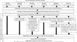
Materials and methods: A total of sixty enamel surfaces of 4 × 5 mm in size were created on both the buccal and lingual sides of thirty extracted wisdom teeth. Remineralization agents were applied to the specimens that were grouped as follows: Group 1, control; Group 2, fluoride varnish (FV); Group 3, P11-4; Group 4, laser; Group 5, laser + FV; and Group 6, laser + P11-4. The fluorescence level was determined with DiagnoDent. The enamel mineral density, area and volume, and caries lesion area and volume were determined with micro-computed tomography (µCT), surface features were evaluated using scanning electron microscopy (SEM), and elemental analysis was performed using energy dispersive x-ray spectroscopy (EDS) .
Results: For specimens treated only with self-assembling peptide P11-4, the caries lesion area (mm2) values were 38.19 and 21.62, and the caries lesion volume (mm3) values were 6.27 and 2.99, respectively for pre- and post-treatment. In combination usage of self-assembling peptide P11-4 and laser, the caries lesion area (mm2) values were 38.39 and 16.91, and the caries lesion volume (mm3) values were 11.15 and 3.64, respectively for pre- and post-treatment. In the application of the P11-4 alone and in combination with laser, there was a statistically significant decrease in DiagnoDent values, an increase in enamel volume(mm3),enamel area(mm2) and mineral density(g/cm3) values and a decrease in caries lesion volume(mm3) and area(mm2) obtained by µCT, and an increase in %Ca and %F values obtained by SEM/EDS analysis (p < 0.05). It was discovered that the samples treated with P11-4 had a considerably higher rise in the Ca/P ratio than the samples treated with FV (p < 0.05). The calcium content increased significantly more when P11-4 application was combined with laser irradiation (p < 0.05).
Conclusions: The combined use of self-assembling peptide P11-4 and laser accelerated the remineralization process and increased the remineralization capacity.
Clinical relevance: FV and P11-4, alone or in combination with laser, can be successfully used as remineralization agents in initial enamel caries.
期刊介绍:
The journal Clinical Oral Investigations is a multidisciplinary, international forum for publication of research from all fields of oral medicine. The journal publishes original scientific articles and invited reviews which provide up-to-date results of basic and clinical studies in oral and maxillofacial science and medicine. The aim is to clarify the relevance of new results to modern practice, for an international readership. Coverage includes maxillofacial and oral surgery, prosthetics and restorative dentistry, operative dentistry, endodontics, periodontology, orthodontics, dental materials science, clinical trials, epidemiology, pedodontics, oral implant, preventive dentistiry, oral pathology, oral basic sciences and more.


 分享
分享
 求助内容:
求助内容: 应助结果提醒方式:
 扫码关注我们
