{"title":"提高肺栓塞的诊断准确性:将低剂量 CT 与 V/Q SPECT 相结合。","authors":"Munassar Dakkam Lasloom, Mohamed Abuzaid","doi":"10.3390/tomography10080096","DOIUrl":null,"url":null,"abstract":"<p><strong>Objective: </strong>This study aimed to retrospectively assess the benefits of combining low-dose computed tomography (LDCT) with ventilation/perfusion single-photon emission computed tomography (V/Q SPECT) for the diagnosis of pulmonary embolism (PE).</p><p><strong>Methods: </strong>A retrospective analysis was performed on 92 patients with suspected PE who underwent V/Q SPECT with ldCT (V/Q SPECT CT) between January 2020 and December 2022 at King Khalid Hospital Najran. Data were collected using the hospital's picture archiving and communication system. Scans were categorized on the basis of perfusion defects, matched or mismatched ventilation, and CT findings. The specificity of V/Q SPECT CT was compared with that of Q SPECT CT.</p><p><strong>Results: </strong>This study included 92 patients (54 females and 38 males; median age, 53 years). The results demonstrated that V/Q SPECT CT had higher specificity (93%) than V/Q SPECT alone (88%). If CT had been used as a ventilation substitute, 21% of patients would have been reported to be positive for PE (8% false-positive), yielding a specificity of 60% for Q SPECT CT. These findings align with the existing literature, although discrepancies in specificity values were noted due to the different study designs and sample sizes.</p><p><strong>Conclusion: </strong>This study highlights the enhanced specificity of V/Q SPECT CT compared to V/Q SPECT and Q SPECT CT alone. Including low-dose CT improves diagnostic accuracy by reducing false positives and providing detailed anatomical information. V/Q SPECT CT offers superior specificity in diagnosing PE compared with V/Q SPECT alone, supporting its use in clinical practice.</p>","PeriodicalId":51330,"journal":{"name":"Tomography","volume":"10 8","pages":"1294-1302"},"PeriodicalIF":2.2000,"publicationDate":"2024-08-16","publicationTypes":"Journal Article","fieldsOfStudy":null,"isOpenAccess":false,"openAccessPdf":"https://www.ncbi.nlm.nih.gov/pmc/articles/PMC11359791/pdf/","citationCount":"0","resultStr":"{\"title\":\"Enhanced Diagnostic Accuracy of Pulmonary Embolism: Integrating Low-Dose CT with V/Q SPECT.\",\"authors\":\"Munassar Dakkam Lasloom, Mohamed Abuzaid\",\"doi\":\"10.3390/tomography10080096\",\"DOIUrl\":null,\"url\":null,\"abstract\":\"<p><strong>Objective: </strong>This study aimed to retrospectively assess the benefits of combining low-dose computed tomography (LDCT) with ventilation/perfusion single-photon emission computed tomography (V/Q SPECT) for the diagnosis of pulmonary embolism (PE).</p><p><strong>Methods: </strong>A retrospective analysis was performed on 92 patients with suspected PE who underwent V/Q SPECT with ldCT (V/Q SPECT CT) between January 2020 and December 2022 at King Khalid Hospital Najran. Data were collected using the hospital's picture archiving and communication system. Scans were categorized on the basis of perfusion defects, matched or mismatched ventilation, and CT findings. The specificity of V/Q SPECT CT was compared with that of Q SPECT CT.</p><p><strong>Results: </strong>This study included 92 patients (54 females and 38 males; median age, 53 years). The results demonstrated that V/Q SPECT CT had higher specificity (93%) than V/Q SPECT alone (88%). If CT had been used as a ventilation substitute, 21% of patients would have been reported to be positive for PE (8% false-positive), yielding a specificity of 60% for Q SPECT CT. These findings align with the existing literature, although discrepancies in specificity values were noted due to the different study designs and sample sizes.</p><p><strong>Conclusion: </strong>This study highlights the enhanced specificity of V/Q SPECT CT compared to V/Q SPECT and Q SPECT CT alone. Including low-dose CT improves diagnostic accuracy by reducing false positives and providing detailed anatomical information. V/Q SPECT CT offers superior specificity in diagnosing PE compared with V/Q SPECT alone, supporting its use in clinical practice.</p>\",\"PeriodicalId\":51330,\"journal\":{\"name\":\"Tomography\",\"volume\":\"10 8\",\"pages\":\"1294-1302\"},\"PeriodicalIF\":2.2000,\"publicationDate\":\"2024-08-16\",\"publicationTypes\":\"Journal Article\",\"fieldsOfStudy\":null,\"isOpenAccess\":false,\"openAccessPdf\":\"https://www.ncbi.nlm.nih.gov/pmc/articles/PMC11359791/pdf/\",\"citationCount\":\"0\",\"resultStr\":null,\"platform\":\"Semanticscholar\",\"paperid\":null,\"PeriodicalName\":\"Tomography\",\"FirstCategoryId\":\"3\",\"ListUrlMain\":\"https://doi.org/10.3390/tomography10080096\",\"RegionNum\":4,\"RegionCategory\":\"医学\",\"ArticlePicture\":[],\"TitleCN\":null,\"AbstractTextCN\":null,\"PMCID\":null,\"EPubDate\":\"\",\"PubModel\":\"\",\"JCR\":\"Q2\",\"JCRName\":\"RADIOLOGY, NUCLEAR MEDICINE & MEDICAL IMAGING\",\"Score\":null,\"Total\":0}","platform":"Semanticscholar","paperid":null,"PeriodicalName":"Tomography","FirstCategoryId":"3","ListUrlMain":"https://doi.org/10.3390/tomography10080096","RegionNum":4,"RegionCategory":"医学","ArticlePicture":[],"TitleCN":null,"AbstractTextCN":null,"PMCID":null,"EPubDate":"","PubModel":"","JCR":"Q2","JCRName":"RADIOLOGY, NUCLEAR MEDICINE & MEDICAL IMAGING","Score":null,"Total":0}
Enhanced Diagnostic Accuracy of Pulmonary Embolism: Integrating Low-Dose CT with V/Q SPECT.
Objective: This study aimed to retrospectively assess the benefits of combining low-dose computed tomography (LDCT) with ventilation/perfusion single-photon emission computed tomography (V/Q SPECT) for the diagnosis of pulmonary embolism (PE).
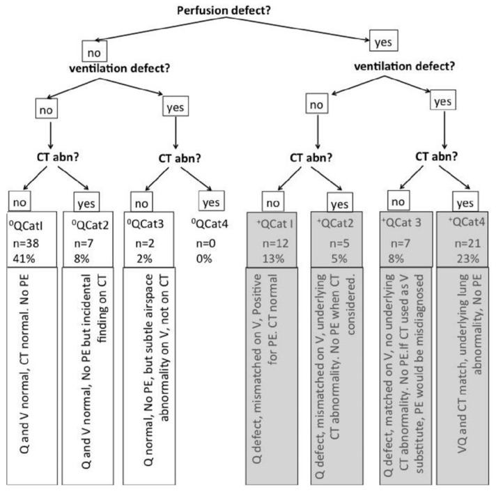
Methods: A retrospective analysis was performed on 92 patients with suspected PE who underwent V/Q SPECT with ldCT (V/Q SPECT CT) between January 2020 and December 2022 at King Khalid Hospital Najran. Data were collected using the hospital's picture archiving and communication system. Scans were categorized on the basis of perfusion defects, matched or mismatched ventilation, and CT findings. The specificity of V/Q SPECT CT was compared with that of Q SPECT CT.
Results: This study included 92 patients (54 females and 38 males; median age, 53 years). The results demonstrated that V/Q SPECT CT had higher specificity (93%) than V/Q SPECT alone (88%). If CT had been used as a ventilation substitute, 21% of patients would have been reported to be positive for PE (8% false-positive), yielding a specificity of 60% for Q SPECT CT. These findings align with the existing literature, although discrepancies in specificity values were noted due to the different study designs and sample sizes.
Conclusion: This study highlights the enhanced specificity of V/Q SPECT CT compared to V/Q SPECT and Q SPECT CT alone. Including low-dose CT improves diagnostic accuracy by reducing false positives and providing detailed anatomical information. V/Q SPECT CT offers superior specificity in diagnosing PE compared with V/Q SPECT alone, supporting its use in clinical practice.
TomographyMedicine-Radiology, Nuclear Medicine and Imaging
CiteScore
2.70
自引率
10.50%
发文量
222
期刊介绍:
TomographyTM publishes basic (technical and pre-clinical) and clinical scientific articles which involve the advancement of imaging technologies. Tomography encompasses studies that use single or multiple imaging modalities including for example CT, US, PET, SPECT, MR and hyperpolarization technologies, as well as optical modalities (i.e. bioluminescence, photoacoustic, endomicroscopy, fiber optic imaging and optical computed tomography) in basic sciences, engineering, preclinical and clinical medicine.
Tomography also welcomes studies involving exploration and refinement of contrast mechanisms and image-derived metrics within and across modalities toward the development of novel imaging probes for image-based feedback and intervention. The use of imaging in biology and medicine provides unparalleled opportunities to noninvasively interrogate tissues to obtain real-time dynamic and quantitative information required for diagnosis and response to interventions and to follow evolving pathological conditions. As multi-modal studies and the complexities of imaging technologies themselves are ever increasing to provide advanced information to scientists and clinicians.
Tomography provides a unique publication venue allowing investigators the opportunity to more precisely communicate integrated findings related to the diverse and heterogeneous features associated with underlying anatomical, physiological, functional, metabolic and molecular genetic activities of normal and diseased tissue. Thus Tomography publishes peer-reviewed articles which involve the broad use of imaging of any tissue and disease type including both preclinical and clinical investigations. In addition, hardware/software along with chemical and molecular probe advances are welcome as they are deemed to significantly contribute towards the long-term goal of improving the overall impact of imaging on scientific and clinical discovery.


 分享
分享
 求助内容:
求助内容: 应助结果提醒方式:
 扫码关注我们
