Eduardo B. Schaustz , José Carlos P. Secco , Julia M. Barroso , Juliana R. Ferreira , Mariana B. Tortelly , Adriana L. Pimentel , Ana Cristina B.S. Figueiredo , Denilson C. Albuquerque , Allan R. Kluser Sales , Paulo H. Rosado de-Castro , Martha V.T. Pinheiro , Olga F. Souza , Emiliano Medei , Ronir R. Luiz , Andréa Silvestre-Sousa , Gabriel C. Camargo , Renata Moll-Bernardes
{"title":"通过磁共振成像检测 COVID-19 幸存者的心脏重塑和炎症情况","authors":"Eduardo B. Schaustz , José Carlos P. Secco , Julia M. Barroso , Juliana R. Ferreira , Mariana B. Tortelly , Adriana L. Pimentel , Ana Cristina B.S. Figueiredo , Denilson C. Albuquerque , Allan R. Kluser Sales , Paulo H. Rosado de-Castro , Martha V.T. Pinheiro , Olga F. Souza , Emiliano Medei , Ronir R. Luiz , Andréa Silvestre-Sousa , Gabriel C. Camargo , Renata Moll-Bernardes","doi":"10.1016/j.ijcha.2024.101499","DOIUrl":null,"url":null,"abstract":"<div><h3>Background</h3><p>Concerns have been raised about cardiac inflammation in patients with long COVID-19, particularly those with myocardial injury during the acute phase of the disease. This study was conducted to examine myopericardial involvement, detected by cardiac magnetic resonance (CMR) imaging in patients hospitalized for COVID-19.</p></div><div><h3>Methods</h3><p>Adult patients hospitalized with COVID-19 who presented myocardial injury or increased D-dimers were enrolled in this prospective study. All patients were invited to undergo CMR imaging examination after discharge. During follow-up, patients with nonischemic myocardial or pericardial involvement detected on the first CMR imaging examination underwent second examinations. CMR imaging findings were compared with those of a control group of healthy patients with no comorbidity.</p></div><div><h3>Results</h3><p>Of 180 included patients, 53 underwent CMR imaging examination. The mean age was 58.4 ± 18.3 years, and 73.6 % were male. Myocardial and pericardial LGE was reported in 43.4 % and 35.8 % of patients, respectively. Nonischemic myocardial or pericardial involvement was reported in 26 (49.1 %) patients. The prevalence of pericardial LGE was associated inversely with the interval between hospital discharge and CMR. COVID-19 survivors had higher end-systolic volume indices (ESVis) and lower left-ventricular ejection fractions than did healthy controls. Seventeen patients underwent follow-up CMR imaging; the end-diastolic volume index, ESVi, and prevalence of pericardial LGE, but not that of nonischemic LGE, were reduced.</p></div><div><h3>Conclusion</h3><p>Among COVID-19 survivors with myocardial injury during the acute phase of the disease, the incidences of nonischemic myocardial and pericardial LGE and CMR imaging–detected signs of cardiac remodeling, partially reversed during follow-up, were high.</p></div>","PeriodicalId":38026,"journal":{"name":"IJC Heart and Vasculature","volume":"54 ","pages":"Article 101499"},"PeriodicalIF":2.9000,"publicationDate":"2024-10-01","publicationTypes":"Journal Article","fieldsOfStudy":null,"isOpenAccess":false,"openAccessPdf":"https://www.sciencedirect.com/science/article/pii/S2352906724001659/pdfft?md5=b4c289219c9b250d73830f5ce40db98e&pid=1-s2.0-S2352906724001659-main.pdf","citationCount":"0","resultStr":"{\"title\":\"Cardiac remodeling and inflammation detected by magnetic resonance imaging in COVID-19 survivors\",\"authors\":\"Eduardo B. Schaustz , José Carlos P. Secco , Julia M. Barroso , Juliana R. Ferreira , Mariana B. Tortelly , Adriana L. Pimentel , Ana Cristina B.S. Figueiredo , Denilson C. Albuquerque , Allan R. Kluser Sales , Paulo H. Rosado de-Castro , Martha V.T. Pinheiro , Olga F. Souza , Emiliano Medei , Ronir R. Luiz , Andréa Silvestre-Sousa , Gabriel C. Camargo , Renata Moll-Bernardes\",\"doi\":\"10.1016/j.ijcha.2024.101499\",\"DOIUrl\":null,\"url\":null,\"abstract\":\"<div><h3>Background</h3><p>Concerns have been raised about cardiac inflammation in patients with long COVID-19, particularly those with myocardial injury during the acute phase of the disease. This study was conducted to examine myopericardial involvement, detected by cardiac magnetic resonance (CMR) imaging in patients hospitalized for COVID-19.</p></div><div><h3>Methods</h3><p>Adult patients hospitalized with COVID-19 who presented myocardial injury or increased D-dimers were enrolled in this prospective study. All patients were invited to undergo CMR imaging examination after discharge. During follow-up, patients with nonischemic myocardial or pericardial involvement detected on the first CMR imaging examination underwent second examinations. CMR imaging findings were compared with those of a control group of healthy patients with no comorbidity.</p></div><div><h3>Results</h3><p>Of 180 included patients, 53 underwent CMR imaging examination. The mean age was 58.4 ± 18.3 years, and 73.6 % were male. Myocardial and pericardial LGE was reported in 43.4 % and 35.8 % of patients, respectively. Nonischemic myocardial or pericardial involvement was reported in 26 (49.1 %) patients. The prevalence of pericardial LGE was associated inversely with the interval between hospital discharge and CMR. COVID-19 survivors had higher end-systolic volume indices (ESVis) and lower left-ventricular ejection fractions than did healthy controls. Seventeen patients underwent follow-up CMR imaging; the end-diastolic volume index, ESVi, and prevalence of pericardial LGE, but not that of nonischemic LGE, were reduced.</p></div><div><h3>Conclusion</h3><p>Among COVID-19 survivors with myocardial injury during the acute phase of the disease, the incidences of nonischemic myocardial and pericardial LGE and CMR imaging–detected signs of cardiac remodeling, partially reversed during follow-up, were high.</p></div>\",\"PeriodicalId\":38026,\"journal\":{\"name\":\"IJC Heart and Vasculature\",\"volume\":\"54 \",\"pages\":\"Article 101499\"},\"PeriodicalIF\":2.9000,\"publicationDate\":\"2024-10-01\",\"publicationTypes\":\"Journal Article\",\"fieldsOfStudy\":null,\"isOpenAccess\":false,\"openAccessPdf\":\"https://www.sciencedirect.com/science/article/pii/S2352906724001659/pdfft?md5=b4c289219c9b250d73830f5ce40db98e&pid=1-s2.0-S2352906724001659-main.pdf\",\"citationCount\":\"0\",\"resultStr\":null,\"platform\":\"Semanticscholar\",\"paperid\":null,\"PeriodicalName\":\"IJC Heart and Vasculature\",\"FirstCategoryId\":\"1085\",\"ListUrlMain\":\"https://www.sciencedirect.com/science/article/pii/S2352906724001659\",\"RegionNum\":0,\"RegionCategory\":null,\"ArticlePicture\":[],\"TitleCN\":null,\"AbstractTextCN\":null,\"PMCID\":null,\"EPubDate\":\"2024/8/27 0:00:00\",\"PubModel\":\"Epub\",\"JCR\":\"Q2\",\"JCRName\":\"CARDIAC & CARDIOVASCULAR SYSTEMS\",\"Score\":null,\"Total\":0}","platform":"Semanticscholar","paperid":null,"PeriodicalName":"IJC Heart and Vasculature","FirstCategoryId":"1085","ListUrlMain":"https://www.sciencedirect.com/science/article/pii/S2352906724001659","RegionNum":0,"RegionCategory":null,"ArticlePicture":[],"TitleCN":null,"AbstractTextCN":null,"PMCID":null,"EPubDate":"2024/8/27 0:00:00","PubModel":"Epub","JCR":"Q2","JCRName":"CARDIAC & CARDIOVASCULAR SYSTEMS","Score":null,"Total":0}
Cardiac remodeling and inflammation detected by magnetic resonance imaging in COVID-19 survivors
Background
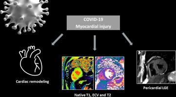
Concerns have been raised about cardiac inflammation in patients with long COVID-19, particularly those with myocardial injury during the acute phase of the disease. This study was conducted to examine myopericardial involvement, detected by cardiac magnetic resonance (CMR) imaging in patients hospitalized for COVID-19.
Methods
Adult patients hospitalized with COVID-19 who presented myocardial injury or increased D-dimers were enrolled in this prospective study. All patients were invited to undergo CMR imaging examination after discharge. During follow-up, patients with nonischemic myocardial or pericardial involvement detected on the first CMR imaging examination underwent second examinations. CMR imaging findings were compared with those of a control group of healthy patients with no comorbidity.
Results
Of 180 included patients, 53 underwent CMR imaging examination. The mean age was 58.4 ± 18.3 years, and 73.6 % were male. Myocardial and pericardial LGE was reported in 43.4 % and 35.8 % of patients, respectively. Nonischemic myocardial or pericardial involvement was reported in 26 (49.1 %) patients. The prevalence of pericardial LGE was associated inversely with the interval between hospital discharge and CMR. COVID-19 survivors had higher end-systolic volume indices (ESVis) and lower left-ventricular ejection fractions than did healthy controls. Seventeen patients underwent follow-up CMR imaging; the end-diastolic volume index, ESVi, and prevalence of pericardial LGE, but not that of nonischemic LGE, were reduced.
Conclusion
Among COVID-19 survivors with myocardial injury during the acute phase of the disease, the incidences of nonischemic myocardial and pericardial LGE and CMR imaging–detected signs of cardiac remodeling, partially reversed during follow-up, were high.
期刊介绍:
IJC Heart & Vasculature is an online-only, open-access journal dedicated to publishing original articles and reviews (also Editorials and Letters to the Editor) which report on structural and functional cardiovascular pathology, with an emphasis on imaging and disease pathophysiology. Articles must be authentic, educational, clinically relevant, and original in their content and scientific approach. IJC Heart & Vasculature requires the highest standards of scientific integrity in order to promote reliable, reproducible and verifiable research findings. All authors are advised to consult the Principles of Ethical Publishing in the International Journal of Cardiology before submitting a manuscript. Submission of a manuscript to this journal gives the publisher the right to publish that paper if it is accepted. Manuscripts may be edited to improve clarity and expression.


 分享
分享
 求助内容:
求助内容: 应助结果提醒方式:
 扫码关注我们
