{"title":"用荧光显微镜观察活体玉米丝(花柱组织)上微生物相互作用的方法","authors":"Michelle E.H. Thompson, Manish N. Raizada","doi":"10.1016/j.mimet.2024.107027","DOIUrl":null,"url":null,"abstract":"<div><p>There is interest in studying microbes that colonize maize silks (style tissue, critical for reproduction) including the fungal pathogen <em>Fusarium graminearum</em> (<em>Fg</em>) and its interactions with the microbiome and biocontrol agents. <em>In planta</em> imaging of these interactions on living silks using confocal fluorescence microscopy would provide key insights. However, newly discovered microbes have unknown effects on human health, and there are regulatory requirements to prevent the release of fluorescently tagged microbes into the environment. Therefore, the microbe infection, colonization, and interaction stages on silks prior to microscopy must be contained. At the same time, silk viability must be maintained and experiments conducted that are biologically relevant (e.g. silks should remain attached to the cob), yet the silk tissue must be accessible to the researcher (i.e. not within husk leaves) and allow for multiple replicates. Here we present methods that meet these five contrasting criteria. We tested these methods using <em>Fg</em> and four silk-derived bacterial endophytes. The endophytes were previously known to have anti-<em>Fg</em> activity in vitro, but <em>in planta</em> observations were lacking. In Method 1, a portion of the tip of a cob was dissected, and silks remained attached to the cob in a Petri dish. The cob was placed on a water agar disc to maintain hydration. DsRed-tagged bacteria and GFP-tagged <em>Fg</em> were inoculated onto the silks and incubated, allowing the two microbes to grow towards one another before staining with propidium iodide for confocal microscopy. A variation of the protocol was presented in Method 2, where detached silk segments were placed directly on water agar where they were inoculated with bacteria and <em>Fg</em> to promote dense colonization, and to allow for many replicates and interventions such as silk wounding. The bacterial endophytes were successfully observed colonizing <em>Fg</em> hyphae, silk trichomes, and entering silks via cut ends and wounds. These protocols can be used to study other silk-associated microbes including several globally important fungal pathogens that enter maize grain through silks.</p></div>","PeriodicalId":16409,"journal":{"name":"Journal of microbiological methods","volume":"225 ","pages":"Article 107027"},"PeriodicalIF":1.9000,"publicationDate":"2024-10-01","publicationTypes":"Journal Article","fieldsOfStudy":null,"isOpenAccess":false,"openAccessPdf":"https://www.sciencedirect.com/science/article/pii/S0167701224001398/pdfft?md5=ceb70988dd3da6a0a097114fe12f9c40&pid=1-s2.0-S0167701224001398-main.pdf","citationCount":"0","resultStr":"{\"title\":\"Protocols to enable fluorescence microscopy of microbial interactions on living maize silks (style tissue)\",\"authors\":\"Michelle E.H. Thompson, Manish N. Raizada\",\"doi\":\"10.1016/j.mimet.2024.107027\",\"DOIUrl\":null,\"url\":null,\"abstract\":\"<div><p>There is interest in studying microbes that colonize maize silks (style tissue, critical for reproduction) including the fungal pathogen <em>Fusarium graminearum</em> (<em>Fg</em>) and its interactions with the microbiome and biocontrol agents. <em>In planta</em> imaging of these interactions on living silks using confocal fluorescence microscopy would provide key insights. However, newly discovered microbes have unknown effects on human health, and there are regulatory requirements to prevent the release of fluorescently tagged microbes into the environment. Therefore, the microbe infection, colonization, and interaction stages on silks prior to microscopy must be contained. At the same time, silk viability must be maintained and experiments conducted that are biologically relevant (e.g. silks should remain attached to the cob), yet the silk tissue must be accessible to the researcher (i.e. not within husk leaves) and allow for multiple replicates. Here we present methods that meet these five contrasting criteria. We tested these methods using <em>Fg</em> and four silk-derived bacterial endophytes. The endophytes were previously known to have anti-<em>Fg</em> activity in vitro, but <em>in planta</em> observations were lacking. In Method 1, a portion of the tip of a cob was dissected, and silks remained attached to the cob in a Petri dish. The cob was placed on a water agar disc to maintain hydration. DsRed-tagged bacteria and GFP-tagged <em>Fg</em> were inoculated onto the silks and incubated, allowing the two microbes to grow towards one another before staining with propidium iodide for confocal microscopy. A variation of the protocol was presented in Method 2, where detached silk segments were placed directly on water agar where they were inoculated with bacteria and <em>Fg</em> to promote dense colonization, and to allow for many replicates and interventions such as silk wounding. The bacterial endophytes were successfully observed colonizing <em>Fg</em> hyphae, silk trichomes, and entering silks via cut ends and wounds. These protocols can be used to study other silk-associated microbes including several globally important fungal pathogens that enter maize grain through silks.</p></div>\",\"PeriodicalId\":16409,\"journal\":{\"name\":\"Journal of microbiological methods\",\"volume\":\"225 \",\"pages\":\"Article 107027\"},\"PeriodicalIF\":1.9000,\"publicationDate\":\"2024-10-01\",\"publicationTypes\":\"Journal Article\",\"fieldsOfStudy\":null,\"isOpenAccess\":false,\"openAccessPdf\":\"https://www.sciencedirect.com/science/article/pii/S0167701224001398/pdfft?md5=ceb70988dd3da6a0a097114fe12f9c40&pid=1-s2.0-S0167701224001398-main.pdf\",\"citationCount\":\"0\",\"resultStr\":null,\"platform\":\"Semanticscholar\",\"paperid\":null,\"PeriodicalName\":\"Journal of microbiological methods\",\"FirstCategoryId\":\"99\",\"ListUrlMain\":\"https://www.sciencedirect.com/science/article/pii/S0167701224001398\",\"RegionNum\":4,\"RegionCategory\":\"生物学\",\"ArticlePicture\":[],\"TitleCN\":null,\"AbstractTextCN\":null,\"PMCID\":null,\"EPubDate\":\"2024/8/29 0:00:00\",\"PubModel\":\"Epub\",\"JCR\":\"Q4\",\"JCRName\":\"BIOCHEMICAL RESEARCH METHODS\",\"Score\":null,\"Total\":0}","platform":"Semanticscholar","paperid":null,"PeriodicalName":"Journal of microbiological methods","FirstCategoryId":"99","ListUrlMain":"https://www.sciencedirect.com/science/article/pii/S0167701224001398","RegionNum":4,"RegionCategory":"生物学","ArticlePicture":[],"TitleCN":null,"AbstractTextCN":null,"PMCID":null,"EPubDate":"2024/8/29 0:00:00","PubModel":"Epub","JCR":"Q4","JCRName":"BIOCHEMICAL RESEARCH METHODS","Score":null,"Total":0}
Protocols to enable fluorescence microscopy of microbial interactions on living maize silks (style tissue)
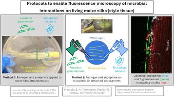
There is interest in studying microbes that colonize maize silks (style tissue, critical for reproduction) including the fungal pathogen Fusarium graminearum (Fg) and its interactions with the microbiome and biocontrol agents. In planta imaging of these interactions on living silks using confocal fluorescence microscopy would provide key insights. However, newly discovered microbes have unknown effects on human health, and there are regulatory requirements to prevent the release of fluorescently tagged microbes into the environment. Therefore, the microbe infection, colonization, and interaction stages on silks prior to microscopy must be contained. At the same time, silk viability must be maintained and experiments conducted that are biologically relevant (e.g. silks should remain attached to the cob), yet the silk tissue must be accessible to the researcher (i.e. not within husk leaves) and allow for multiple replicates. Here we present methods that meet these five contrasting criteria. We tested these methods using Fg and four silk-derived bacterial endophytes. The endophytes were previously known to have anti-Fg activity in vitro, but in planta observations were lacking. In Method 1, a portion of the tip of a cob was dissected, and silks remained attached to the cob in a Petri dish. The cob was placed on a water agar disc to maintain hydration. DsRed-tagged bacteria and GFP-tagged Fg were inoculated onto the silks and incubated, allowing the two microbes to grow towards one another before staining with propidium iodide for confocal microscopy. A variation of the protocol was presented in Method 2, where detached silk segments were placed directly on water agar where they were inoculated with bacteria and Fg to promote dense colonization, and to allow for many replicates and interventions such as silk wounding. The bacterial endophytes were successfully observed colonizing Fg hyphae, silk trichomes, and entering silks via cut ends and wounds. These protocols can be used to study other silk-associated microbes including several globally important fungal pathogens that enter maize grain through silks.
期刊介绍:
The Journal of Microbiological Methods publishes scholarly and original articles, notes and review articles. These articles must include novel and/or state-of-the-art methods, or significant improvements to existing methods. Novel and innovative applications of current methods that are validated and useful will also be published. JMM strives for scholarship, innovation and excellence. This demands scientific rigour, the best available methods and technologies, correctly replicated experiments/tests, the inclusion of proper controls, calibrations, and the correct statistical analysis. The presentation of the data must support the interpretation of the method/approach.
All aspects of microbiology are covered, except virology. These include agricultural microbiology, applied and environmental microbiology, bioassays, bioinformatics, biotechnology, biochemical microbiology, clinical microbiology, diagnostics, food monitoring and quality control microbiology, microbial genetics and genomics, geomicrobiology, microbiome methods regardless of habitat, high through-put sequencing methods and analysis, microbial pathogenesis and host responses, metabolomics, metagenomics, metaproteomics, microbial ecology and diversity, microbial physiology, microbial ultra-structure, microscopic and imaging methods, molecular microbiology, mycology, novel mathematical microbiology and modelling, parasitology, plant-microbe interactions, protein markers/profiles, proteomics, pyrosequencing, public health microbiology, radioisotopes applied to microbiology, robotics applied to microbiological methods,rumen microbiology, microbiological methods for space missions and extreme environments, sampling methods and samplers, soil and sediment microbiology, transcriptomics, veterinary microbiology, sero-diagnostics and typing/identification.


 分享
分享
 求助内容:
求助内容: 应助结果提醒方式:
 扫码关注我们
