Young Woo Kim, Simon Biggs, Elizabeth Claridge Mackonis
{"title":"基于人工智能的多种自动轮廓系统在风险器官(OARs)划定中的性能研究。","authors":"Young Woo Kim, Simon Biggs, Elizabeth Claridge Mackonis","doi":"10.1007/s13246-024-01434-9","DOIUrl":null,"url":null,"abstract":"<p><p>Manual contouring of organs at risk (OAR) is time-consuming and subject to inter-observer variability. AI-based auto-contouring is proposed as a solution to these problems if it can produce clinically acceptable results. This study investigated the performance of multiple AI-based auto-contouring systems in different OAR segmentations. The auto-contouring was performed using seven different AI-based segmentation systems (Radiotherapy AI, Limbus AI version 1.5 and 1.6, Therapanacea, MIM, Siemens AI-Rad Companion and RadFormation) on a total of 42 clinical cases with varying anatomical sites. Volumetric and surface dice similarity coefficients and maximum Hausdorff distance (HD) between the expert's contours and automated contours were calculated to evaluate their performance. Radiotherapy AI has shown better performance than other software in most tested structures considered in the head and neck, and brain cases. No specific software had shown overall superior performance over other software in lung, breast, pelvis and abdomen cases. Each tested AI system was able to produce comparable contours to the experts' contours of organs at risk which can potentially be used for clinical use. A reduced performance of AI systems in the case of small and complex anatomical structures was found and reported, showing that it is still essential to review each contour produced by AI systems for clinical uses. This study has also demonstrated a method of comparing contouring software options which could be replicated in clinics or used for ongoing quality assurance of purchased systems.</p>","PeriodicalId":48490,"journal":{"name":"Physical and Engineering Sciences in Medicine","volume":" ","pages":"1123-1140"},"PeriodicalIF":2.4000,"publicationDate":"2024-09-01","publicationTypes":"Journal Article","fieldsOfStudy":null,"isOpenAccess":false,"openAccessPdf":"https://www.ncbi.nlm.nih.gov/pmc/articles/PMC11408550/pdf/","citationCount":"0","resultStr":"{\"title\":\"Investigation on performance of multiple AI-based auto-contouring systems in organs at risks (OARs) delineation.\",\"authors\":\"Young Woo Kim, Simon Biggs, Elizabeth Claridge Mackonis\",\"doi\":\"10.1007/s13246-024-01434-9\",\"DOIUrl\":null,\"url\":null,\"abstract\":\"<p><p>Manual contouring of organs at risk (OAR) is time-consuming and subject to inter-observer variability. AI-based auto-contouring is proposed as a solution to these problems if it can produce clinically acceptable results. This study investigated the performance of multiple AI-based auto-contouring systems in different OAR segmentations. The auto-contouring was performed using seven different AI-based segmentation systems (Radiotherapy AI, Limbus AI version 1.5 and 1.6, Therapanacea, MIM, Siemens AI-Rad Companion and RadFormation) on a total of 42 clinical cases with varying anatomical sites. Volumetric and surface dice similarity coefficients and maximum Hausdorff distance (HD) between the expert's contours and automated contours were calculated to evaluate their performance. Radiotherapy AI has shown better performance than other software in most tested structures considered in the head and neck, and brain cases. No specific software had shown overall superior performance over other software in lung, breast, pelvis and abdomen cases. Each tested AI system was able to produce comparable contours to the experts' contours of organs at risk which can potentially be used for clinical use. A reduced performance of AI systems in the case of small and complex anatomical structures was found and reported, showing that it is still essential to review each contour produced by AI systems for clinical uses. This study has also demonstrated a method of comparing contouring software options which could be replicated in clinics or used for ongoing quality assurance of purchased systems.</p>\",\"PeriodicalId\":48490,\"journal\":{\"name\":\"Physical and Engineering Sciences in Medicine\",\"volume\":\" \",\"pages\":\"1123-1140\"},\"PeriodicalIF\":2.4000,\"publicationDate\":\"2024-09-01\",\"publicationTypes\":\"Journal Article\",\"fieldsOfStudy\":null,\"isOpenAccess\":false,\"openAccessPdf\":\"https://www.ncbi.nlm.nih.gov/pmc/articles/PMC11408550/pdf/\",\"citationCount\":\"0\",\"resultStr\":null,\"platform\":\"Semanticscholar\",\"paperid\":null,\"PeriodicalName\":\"Physical and Engineering Sciences in Medicine\",\"FirstCategoryId\":\"3\",\"ListUrlMain\":\"https://doi.org/10.1007/s13246-024-01434-9\",\"RegionNum\":4,\"RegionCategory\":\"医学\",\"ArticlePicture\":[],\"TitleCN\":null,\"AbstractTextCN\":null,\"PMCID\":null,\"EPubDate\":\"2024/9/2 0:00:00\",\"PubModel\":\"Epub\",\"JCR\":\"Q3\",\"JCRName\":\"ENGINEERING, BIOMEDICAL\",\"Score\":null,\"Total\":0}","platform":"Semanticscholar","paperid":null,"PeriodicalName":"Physical and Engineering Sciences in Medicine","FirstCategoryId":"3","ListUrlMain":"https://doi.org/10.1007/s13246-024-01434-9","RegionNum":4,"RegionCategory":"医学","ArticlePicture":[],"TitleCN":null,"AbstractTextCN":null,"PMCID":null,"EPubDate":"2024/9/2 0:00:00","PubModel":"Epub","JCR":"Q3","JCRName":"ENGINEERING, BIOMEDICAL","Score":null,"Total":0}
Investigation on performance of multiple AI-based auto-contouring systems in organs at risks (OARs) delineation.
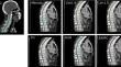
Manual contouring of organs at risk (OAR) is time-consuming and subject to inter-observer variability. AI-based auto-contouring is proposed as a solution to these problems if it can produce clinically acceptable results. This study investigated the performance of multiple AI-based auto-contouring systems in different OAR segmentations. The auto-contouring was performed using seven different AI-based segmentation systems (Radiotherapy AI, Limbus AI version 1.5 and 1.6, Therapanacea, MIM, Siemens AI-Rad Companion and RadFormation) on a total of 42 clinical cases with varying anatomical sites. Volumetric and surface dice similarity coefficients and maximum Hausdorff distance (HD) between the expert's contours and automated contours were calculated to evaluate their performance. Radiotherapy AI has shown better performance than other software in most tested structures considered in the head and neck, and brain cases. No specific software had shown overall superior performance over other software in lung, breast, pelvis and abdomen cases. Each tested AI system was able to produce comparable contours to the experts' contours of organs at risk which can potentially be used for clinical use. A reduced performance of AI systems in the case of small and complex anatomical structures was found and reported, showing that it is still essential to review each contour produced by AI systems for clinical uses. This study has also demonstrated a method of comparing contouring software options which could be replicated in clinics or used for ongoing quality assurance of purchased systems.


 分享
分享
 求助内容:
求助内容: 应助结果提醒方式:
 扫码关注我们
