{"title":"活细胞成像和 CLEM 揭示了 ACTN4 依赖性皱边薄片的存在,它是一种新型的细胞迁移模式。","authors":"Haruka Morishita , Katsuhisa Kawai , Youhei Egami , Kazufumi Honda , Nobukazu Araki","doi":"10.1016/j.yexcr.2024.114232","DOIUrl":null,"url":null,"abstract":"<div><p>α-Actinin-4 (ACTN4) expression levels are correlated with the invasive and metastatic potential of cancer cells; however, the underlying mechanism remains unclear. Here, we identified ACTN4-localized ruffle-edge lamellipodia using live-cell imaging and correlative light and electron microscopy (CLEM). BSC-1 cells expressing EGFP-ACTN4 showed that ACTN4 was most abundant in the leading edges of lamellipodia, although it was also present in stress fibers and focal adhesions. ACTN4 localization in lamellipodia was markedly diminished by phosphoinositide 3-kinase inhibition, whereas its localization in stress fibers and focal adhesions remained. Furthermore, overexpression of ACTN4, but not ACTN1, promoted lamellipodial formation. Live-cell analysis demonstrated that ACTN4-enriched lamellipodia are highly dynamic and associated with cell migration. CLEM revealed that ACTN4-enriched lamellipodia exhibit a characteristic morphology of multilayered ruffle-edges that differs from canonical flat lamellipodia. Similar ruffle-edge lamellipodia were observed in A549 and MDA-MB-231 invasive cancer cells. ACTN4 knockdown suppressed the formation of ruffle-edge lamellipodia and cell migration during wound healing in A549 monolayer cultures. Additionally, membrane-type 1 matrix metalloproteinase was observed in the membrane ruffles, suggesting that ruffle-edge lamellipodia have the ability to degrade the extracellular matrix and may contribute to active cell migration/invasion in certain cancer cell types.</p></div>","PeriodicalId":12227,"journal":{"name":"Experimental cell research","volume":"442 2","pages":"Article 114232"},"PeriodicalIF":3.5000,"publicationDate":"2024-10-01","publicationTypes":"Journal Article","fieldsOfStudy":null,"isOpenAccess":false,"openAccessPdf":"","citationCount":"0","resultStr":"{\"title\":\"Live-cell imaging and CLEM reveal the existence of ACTN4-dependent ruffle-edge lamellipodia acting as a novel mode of cell migration\",\"authors\":\"Haruka Morishita , Katsuhisa Kawai , Youhei Egami , Kazufumi Honda , Nobukazu Araki\",\"doi\":\"10.1016/j.yexcr.2024.114232\",\"DOIUrl\":null,\"url\":null,\"abstract\":\"<div><p>α-Actinin-4 (ACTN4) expression levels are correlated with the invasive and metastatic potential of cancer cells; however, the underlying mechanism remains unclear. Here, we identified ACTN4-localized ruffle-edge lamellipodia using live-cell imaging and correlative light and electron microscopy (CLEM). BSC-1 cells expressing EGFP-ACTN4 showed that ACTN4 was most abundant in the leading edges of lamellipodia, although it was also present in stress fibers and focal adhesions. ACTN4 localization in lamellipodia was markedly diminished by phosphoinositide 3-kinase inhibition, whereas its localization in stress fibers and focal adhesions remained. Furthermore, overexpression of ACTN4, but not ACTN1, promoted lamellipodial formation. Live-cell analysis demonstrated that ACTN4-enriched lamellipodia are highly dynamic and associated with cell migration. CLEM revealed that ACTN4-enriched lamellipodia exhibit a characteristic morphology of multilayered ruffle-edges that differs from canonical flat lamellipodia. Similar ruffle-edge lamellipodia were observed in A549 and MDA-MB-231 invasive cancer cells. ACTN4 knockdown suppressed the formation of ruffle-edge lamellipodia and cell migration during wound healing in A549 monolayer cultures. Additionally, membrane-type 1 matrix metalloproteinase was observed in the membrane ruffles, suggesting that ruffle-edge lamellipodia have the ability to degrade the extracellular matrix and may contribute to active cell migration/invasion in certain cancer cell types.</p></div>\",\"PeriodicalId\":12227,\"journal\":{\"name\":\"Experimental cell research\",\"volume\":\"442 2\",\"pages\":\"Article 114232\"},\"PeriodicalIF\":3.5000,\"publicationDate\":\"2024-10-01\",\"publicationTypes\":\"Journal Article\",\"fieldsOfStudy\":null,\"isOpenAccess\":false,\"openAccessPdf\":\"\",\"citationCount\":\"0\",\"resultStr\":null,\"platform\":\"Semanticscholar\",\"paperid\":null,\"PeriodicalName\":\"Experimental cell research\",\"FirstCategoryId\":\"3\",\"ListUrlMain\":\"https://www.sciencedirect.com/science/article/pii/S0014482724003239\",\"RegionNum\":3,\"RegionCategory\":\"生物学\",\"ArticlePicture\":[],\"TitleCN\":null,\"AbstractTextCN\":null,\"PMCID\":null,\"EPubDate\":\"2024/9/1 0:00:00\",\"PubModel\":\"Epub\",\"JCR\":\"Q3\",\"JCRName\":\"CELL BIOLOGY\",\"Score\":null,\"Total\":0}","platform":"Semanticscholar","paperid":null,"PeriodicalName":"Experimental cell research","FirstCategoryId":"3","ListUrlMain":"https://www.sciencedirect.com/science/article/pii/S0014482724003239","RegionNum":3,"RegionCategory":"生物学","ArticlePicture":[],"TitleCN":null,"AbstractTextCN":null,"PMCID":null,"EPubDate":"2024/9/1 0:00:00","PubModel":"Epub","JCR":"Q3","JCRName":"CELL BIOLOGY","Score":null,"Total":0}
Live-cell imaging and CLEM reveal the existence of ACTN4-dependent ruffle-edge lamellipodia acting as a novel mode of cell migration
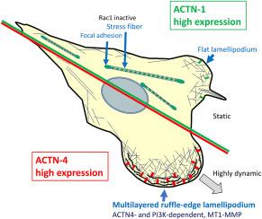
α-Actinin-4 (ACTN4) expression levels are correlated with the invasive and metastatic potential of cancer cells; however, the underlying mechanism remains unclear. Here, we identified ACTN4-localized ruffle-edge lamellipodia using live-cell imaging and correlative light and electron microscopy (CLEM). BSC-1 cells expressing EGFP-ACTN4 showed that ACTN4 was most abundant in the leading edges of lamellipodia, although it was also present in stress fibers and focal adhesions. ACTN4 localization in lamellipodia was markedly diminished by phosphoinositide 3-kinase inhibition, whereas its localization in stress fibers and focal adhesions remained. Furthermore, overexpression of ACTN4, but not ACTN1, promoted lamellipodial formation. Live-cell analysis demonstrated that ACTN4-enriched lamellipodia are highly dynamic and associated with cell migration. CLEM revealed that ACTN4-enriched lamellipodia exhibit a characteristic morphology of multilayered ruffle-edges that differs from canonical flat lamellipodia. Similar ruffle-edge lamellipodia were observed in A549 and MDA-MB-231 invasive cancer cells. ACTN4 knockdown suppressed the formation of ruffle-edge lamellipodia and cell migration during wound healing in A549 monolayer cultures. Additionally, membrane-type 1 matrix metalloproteinase was observed in the membrane ruffles, suggesting that ruffle-edge lamellipodia have the ability to degrade the extracellular matrix and may contribute to active cell migration/invasion in certain cancer cell types.
期刊介绍:
Our scope includes but is not limited to areas such as: Chromosome biology; Chromatin and epigenetics; DNA repair; Gene regulation; Nuclear import-export; RNA processing; Non-coding RNAs; Organelle biology; The cytoskeleton; Intracellular trafficking; Cell-cell and cell-matrix interactions; Cell motility and migration; Cell proliferation; Cellular differentiation; Signal transduction; Programmed cell death.


 分享
分享
 求助内容:
求助内容: 应助结果提醒方式:
 扫码关注我们
