Eunkyung Kim, Seo Jung Yun, Byung-Mo Oh, Han Gil Seo
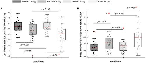
{"title":"经颅直流电刺激左侧背外侧前额叶皮层的电场幅度对内在功能连接性变化的影响:随机交叉研究》。","authors":"Eunkyung Kim, Seo Jung Yun, Byung-Mo Oh, Han Gil Seo","doi":"10.1002/jnr.25378","DOIUrl":null,"url":null,"abstract":"<p>This study investigated whether the electric field magnitude (E-field) delivered to the left dorsolateral prefrontal cortex (L-DLPFC) changes resting-state brain activity and the L-DLPFC resting-state functional connectivity (rsFC), given the variability in tDCS response and lack of understanding of how rsFC changes. Twenty-one healthy participants received either 2 mA anodal or sham tDCS targeting the L-DLPFC for 10 min. Brain imaging was conducted before and after stimulation. The fractional amplitude of low-frequency fluctuation (fALFF), reflecting resting brain activity, and the L-DLPFC rsFC were analyzed to investigate the main effect of tDCS, main effect of time, and interaction effects. The E-field was estimated by modeling tDCS-induced individual electric fields and correlated with fALFF and L-DLPFC rsFC. Anodal tDCS increased fALFF in the left rostral middle frontal area and decreased fALFF in the midline frontal area (FWE <i>p</i> < 0.050), whereas sham induced no changes. Overall rsFC decreased after sham (positive and negative connectivity, <i>p</i> = 0.001 and 0.020, respectively), with modest and nonsignificant changes after anodal tDCS (<i>p</i> = 0.063 and 0.069, respectively). No significant differences in local rsFC were observed among the conditions. Correlations were observed between the E-field and rsFC changes in the L-DLPFC (<i>r</i> = 0.385, <i>p</i> = 0.115), left inferior parietal area (<i>r</i> = 0.495, <i>p</i> = 0.037), and right lateral visual area (<i>r</i> = 0.683, <i>p</i> = 0.002). Single-session tDCS induced resting brain activity changes and may help maintain overall rsFC. The E-field in the L-DLPFC is associated with rsFC changes in both proximal and distally connected brain regions to the L-DLPFC.</p>","PeriodicalId":16490,"journal":{"name":"Journal of Neuroscience Research","volume":"102 9","pages":""},"PeriodicalIF":3.4000,"publicationDate":"2024-09-03","publicationTypes":"Journal Article","fieldsOfStudy":null,"isOpenAccess":false,"openAccessPdf":"https://onlinelibrary.wiley.com/doi/epdf/10.1002/jnr.25378","citationCount":"0","resultStr":"{\"title\":\"Impact of Electric Field Magnitude in the Left Dorsolateral Prefrontal Cortex on Changes in Intrinsic Functional Connectivity Using Transcranial Direct Current Stimulation: A Randomized Crossover Study\",\"authors\":\"Eunkyung Kim, Seo Jung Yun, Byung-Mo Oh, Han Gil Seo\",\"doi\":\"10.1002/jnr.25378\",\"DOIUrl\":null,\"url\":null,\"abstract\":\"<p>This study investigated whether the electric field magnitude (E-field) delivered to the left dorsolateral prefrontal cortex (L-DLPFC) changes resting-state brain activity and the L-DLPFC resting-state functional connectivity (rsFC), given the variability in tDCS response and lack of understanding of how rsFC changes. Twenty-one healthy participants received either 2 mA anodal or sham tDCS targeting the L-DLPFC for 10 min. Brain imaging was conducted before and after stimulation. The fractional amplitude of low-frequency fluctuation (fALFF), reflecting resting brain activity, and the L-DLPFC rsFC were analyzed to investigate the main effect of tDCS, main effect of time, and interaction effects. The E-field was estimated by modeling tDCS-induced individual electric fields and correlated with fALFF and L-DLPFC rsFC. Anodal tDCS increased fALFF in the left rostral middle frontal area and decreased fALFF in the midline frontal area (FWE <i>p</i> < 0.050), whereas sham induced no changes. Overall rsFC decreased after sham (positive and negative connectivity, <i>p</i> = 0.001 and 0.020, respectively), with modest and nonsignificant changes after anodal tDCS (<i>p</i> = 0.063 and 0.069, respectively). No significant differences in local rsFC were observed among the conditions. Correlations were observed between the E-field and rsFC changes in the L-DLPFC (<i>r</i> = 0.385, <i>p</i> = 0.115), left inferior parietal area (<i>r</i> = 0.495, <i>p</i> = 0.037), and right lateral visual area (<i>r</i> = 0.683, <i>p</i> = 0.002). Single-session tDCS induced resting brain activity changes and may help maintain overall rsFC. The E-field in the L-DLPFC is associated with rsFC changes in both proximal and distally connected brain regions to the L-DLPFC.</p>\",\"PeriodicalId\":16490,\"journal\":{\"name\":\"Journal of Neuroscience Research\",\"volume\":\"102 9\",\"pages\":\"\"},\"PeriodicalIF\":3.4000,\"publicationDate\":\"2024-09-03\",\"publicationTypes\":\"Journal Article\",\"fieldsOfStudy\":null,\"isOpenAccess\":false,\"openAccessPdf\":\"https://onlinelibrary.wiley.com/doi/epdf/10.1002/jnr.25378\",\"citationCount\":\"0\",\"resultStr\":null,\"platform\":\"Semanticscholar\",\"paperid\":null,\"PeriodicalName\":\"Journal of Neuroscience Research\",\"FirstCategoryId\":\"3\",\"ListUrlMain\":\"https://onlinelibrary.wiley.com/doi/10.1002/jnr.25378\",\"RegionNum\":3,\"RegionCategory\":\"医学\",\"ArticlePicture\":[],\"TitleCN\":null,\"AbstractTextCN\":null,\"PMCID\":null,\"EPubDate\":\"\",\"PubModel\":\"\",\"JCR\":\"Q2\",\"JCRName\":\"NEUROSCIENCES\",\"Score\":null,\"Total\":0}","platform":"Semanticscholar","paperid":null,"PeriodicalName":"Journal of Neuroscience Research","FirstCategoryId":"3","ListUrlMain":"https://onlinelibrary.wiley.com/doi/10.1002/jnr.25378","RegionNum":3,"RegionCategory":"医学","ArticlePicture":[],"TitleCN":null,"AbstractTextCN":null,"PMCID":null,"EPubDate":"","PubModel":"","JCR":"Q2","JCRName":"NEUROSCIENCES","Score":null,"Total":0}
Impact of Electric Field Magnitude in the Left Dorsolateral Prefrontal Cortex on Changes in Intrinsic Functional Connectivity Using Transcranial Direct Current Stimulation: A Randomized Crossover Study
This study investigated whether the electric field magnitude (E-field) delivered to the left dorsolateral prefrontal cortex (L-DLPFC) changes resting-state brain activity and the L-DLPFC resting-state functional connectivity (rsFC), given the variability in tDCS response and lack of understanding of how rsFC changes. Twenty-one healthy participants received either 2 mA anodal or sham tDCS targeting the L-DLPFC for 10 min. Brain imaging was conducted before and after stimulation. The fractional amplitude of low-frequency fluctuation (fALFF), reflecting resting brain activity, and the L-DLPFC rsFC were analyzed to investigate the main effect of tDCS, main effect of time, and interaction effects. The E-field was estimated by modeling tDCS-induced individual electric fields and correlated with fALFF and L-DLPFC rsFC. Anodal tDCS increased fALFF in the left rostral middle frontal area and decreased fALFF in the midline frontal area (FWE p < 0.050), whereas sham induced no changes. Overall rsFC decreased after sham (positive and negative connectivity, p = 0.001 and 0.020, respectively), with modest and nonsignificant changes after anodal tDCS (p = 0.063 and 0.069, respectively). No significant differences in local rsFC were observed among the conditions. Correlations were observed between the E-field and rsFC changes in the L-DLPFC (r = 0.385, p = 0.115), left inferior parietal area (r = 0.495, p = 0.037), and right lateral visual area (r = 0.683, p = 0.002). Single-session tDCS induced resting brain activity changes and may help maintain overall rsFC. The E-field in the L-DLPFC is associated with rsFC changes in both proximal and distally connected brain regions to the L-DLPFC.
期刊介绍:
The Journal of Neuroscience Research (JNR) publishes novel research results that will advance our understanding of the development, function and pathophysiology of the nervous system, using molecular, cellular, systems, and translational approaches. JNR covers both basic research and clinical aspects of neurology, neuropathology, psychiatry or psychology.
The journal focuses on uncovering the intricacies of brain structure and function. Research published in JNR covers all species from invertebrates to humans, and the reports inform the readers about the function and organization of the nervous system, with emphasis on how disease modifies the function and organization.


 分享
分享
 求助内容:
求助内容: 应助结果提醒方式:
 扫码关注我们
