{"title":"中颅窝 Mullan 三角区或前内侧三角区:尸体研究及其外科重要性。","authors":"Ariyanachi Kaliappan, Rohini Motwani, Mrudula Chandrupatla, Apurba Patra","doi":"10.1007/s00276-024-03475-x","DOIUrl":null,"url":null,"abstract":"<p><strong>Background: </strong>Surgical approaches to the cavernous sinus (CS) and middle cranial fossa (MCF) can be challenging, particularly for young neurosurgeons. The anteromedial (Mullan's) triangle is a triangle by the side of the CS and constitutes part of the floor of the MCF. The contents include the sphenoid sinus, superior ophthalmic vein, and sixth cranial nerve. The literature contains very little research that has precisely defined and measured the anteromedial triangle while considering anatomical variances minimally.</p><p><strong>Methodology: </strong>The present study was conducted on the skulls of 25 adult human cadavers which were dissected to expose the anteromedial (Mullan's) triangle on both sides. After precisely defining the triangle on each side, measurements of the three borders were taken, and using Heron's formula, the area of each triangle was calculated.</p><p><strong>Results: </strong>On average, the length of the medial border was 12.5 (+ 3.1 mm); the length of the lateral border was 9.9 (+ 3.1 mm); the length of the base was 10.75 (+ 2.4 mm) and the area of the anteromedial triangle was 43.9 (+ 15.06 mm<sup>2</sup>).</p><p><strong>Conclusion: </strong>Precise anatomical knowledge of the Mullan's triangle enables the treatment of disorders in often deformed anatomy or difficult-to-access structures. That is the reason it is important to gain a thorough understanding of the surgical anatomy and to adopt a safe procedure.</p>","PeriodicalId":49461,"journal":{"name":"Surgical and Radiologic Anatomy","volume":" ","pages":"1761-1767"},"PeriodicalIF":1.2000,"publicationDate":"2024-11-01","publicationTypes":"Journal Article","fieldsOfStudy":null,"isOpenAccess":false,"openAccessPdf":"","citationCount":"0","resultStr":"{\"title\":\"Mullan's triangle or anteromedial triangle of the middle cranial fossa: a cadaveric study with its surgical importance.\",\"authors\":\"Ariyanachi Kaliappan, Rohini Motwani, Mrudula Chandrupatla, Apurba Patra\",\"doi\":\"10.1007/s00276-024-03475-x\",\"DOIUrl\":null,\"url\":null,\"abstract\":\"<p><strong>Background: </strong>Surgical approaches to the cavernous sinus (CS) and middle cranial fossa (MCF) can be challenging, particularly for young neurosurgeons. The anteromedial (Mullan's) triangle is a triangle by the side of the CS and constitutes part of the floor of the MCF. The contents include the sphenoid sinus, superior ophthalmic vein, and sixth cranial nerve. The literature contains very little research that has precisely defined and measured the anteromedial triangle while considering anatomical variances minimally.</p><p><strong>Methodology: </strong>The present study was conducted on the skulls of 25 adult human cadavers which were dissected to expose the anteromedial (Mullan's) triangle on both sides. After precisely defining the triangle on each side, measurements of the three borders were taken, and using Heron's formula, the area of each triangle was calculated.</p><p><strong>Results: </strong>On average, the length of the medial border was 12.5 (+ 3.1 mm); the length of the lateral border was 9.9 (+ 3.1 mm); the length of the base was 10.75 (+ 2.4 mm) and the area of the anteromedial triangle was 43.9 (+ 15.06 mm<sup>2</sup>).</p><p><strong>Conclusion: </strong>Precise anatomical knowledge of the Mullan's triangle enables the treatment of disorders in often deformed anatomy or difficult-to-access structures. That is the reason it is important to gain a thorough understanding of the surgical anatomy and to adopt a safe procedure.</p>\",\"PeriodicalId\":49461,\"journal\":{\"name\":\"Surgical and Radiologic Anatomy\",\"volume\":\" \",\"pages\":\"1761-1767\"},\"PeriodicalIF\":1.2000,\"publicationDate\":\"2024-11-01\",\"publicationTypes\":\"Journal Article\",\"fieldsOfStudy\":null,\"isOpenAccess\":false,\"openAccessPdf\":\"\",\"citationCount\":\"0\",\"resultStr\":null,\"platform\":\"Semanticscholar\",\"paperid\":null,\"PeriodicalName\":\"Surgical and Radiologic Anatomy\",\"FirstCategoryId\":\"3\",\"ListUrlMain\":\"https://doi.org/10.1007/s00276-024-03475-x\",\"RegionNum\":4,\"RegionCategory\":\"医学\",\"ArticlePicture\":[],\"TitleCN\":null,\"AbstractTextCN\":null,\"PMCID\":null,\"EPubDate\":\"2024/9/3 0:00:00\",\"PubModel\":\"Epub\",\"JCR\":\"Q2\",\"JCRName\":\"Medicine\",\"Score\":null,\"Total\":0}","platform":"Semanticscholar","paperid":null,"PeriodicalName":"Surgical and Radiologic Anatomy","FirstCategoryId":"3","ListUrlMain":"https://doi.org/10.1007/s00276-024-03475-x","RegionNum":4,"RegionCategory":"医学","ArticlePicture":[],"TitleCN":null,"AbstractTextCN":null,"PMCID":null,"EPubDate":"2024/9/3 0:00:00","PubModel":"Epub","JCR":"Q2","JCRName":"Medicine","Score":null,"Total":0}
Mullan's triangle or anteromedial triangle of the middle cranial fossa: a cadaveric study with its surgical importance.
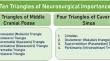
Background: Surgical approaches to the cavernous sinus (CS) and middle cranial fossa (MCF) can be challenging, particularly for young neurosurgeons. The anteromedial (Mullan's) triangle is a triangle by the side of the CS and constitutes part of the floor of the MCF. The contents include the sphenoid sinus, superior ophthalmic vein, and sixth cranial nerve. The literature contains very little research that has precisely defined and measured the anteromedial triangle while considering anatomical variances minimally.
Methodology: The present study was conducted on the skulls of 25 adult human cadavers which were dissected to expose the anteromedial (Mullan's) triangle on both sides. After precisely defining the triangle on each side, measurements of the three borders were taken, and using Heron's formula, the area of each triangle was calculated.
Results: On average, the length of the medial border was 12.5 (+ 3.1 mm); the length of the lateral border was 9.9 (+ 3.1 mm); the length of the base was 10.75 (+ 2.4 mm) and the area of the anteromedial triangle was 43.9 (+ 15.06 mm2).
Conclusion: Precise anatomical knowledge of the Mullan's triangle enables the treatment of disorders in often deformed anatomy or difficult-to-access structures. That is the reason it is important to gain a thorough understanding of the surgical anatomy and to adopt a safe procedure.
期刊介绍:
Anatomy is a morphological science which cannot fail to interest the clinician. The practical application of anatomical research to clinical problems necessitates special adaptation and selectivity in choosing from numerous international works. Although there is a tendency to believe that meaningful advances in anatomy are unlikely, constant revision is necessary. Surgical and Radiologic Anatomy, the first international journal of Clinical anatomy has been created in this spirit.
Its goal is to serve clinicians, regardless of speciality-physicians, surgeons, radiologists or other specialists-as an indispensable aid with which they can improve their knowledge of anatomy. Each issue includes: Original papers, review articles, articles on the anatomical bases of medical, surgical and radiological techniques, articles of normal radiologic anatomy, brief reviews of anatomical publications of clinical interest.
Particular attention is given to high quality illustrations, which are indispensable for a better understanding of anatomical problems.
Surgical and Radiologic Anatomy is a journal written by anatomists for clinicians with a special interest in anatomy.


 分享
分享
 求助内容:
求助内容: 应助结果提醒方式:
 扫码关注我们
