Shiwei Huang, Troy Lund, Paul Orchard, Ashish Gupta, David Nascene
{"title":"赫勒综合征患者吉布斯畸形的发病率和自然史。","authors":"Shiwei Huang, Troy Lund, Paul Orchard, Ashish Gupta, David Nascene","doi":"10.1007/s00234-024-03462-4","DOIUrl":null,"url":null,"abstract":"<p><strong>Introduction: </strong>Gibbus deformity has been documented as a common musculoskeletal abnormality in mucopolysaccharidosis type I (Hurler syndrome, MPS IH), and its recognition often leads to the diagnosis of MPS IH. While the incidence has been described, the progression of gibbus deformities is not well known. Here we describe the natural history of gibbus deformity in a single center patient population using serial spinal MRI scans.</p><p><strong>Methods: </strong>All spinal MRI scans in MPS IH patients were retrospectively reviewed. The presence, spinal location, and angulation of the gibbus deformities were collected. The angles between the superior endplate of the superior normal vertebral body and the inferior endplate of the inferior normal vertebral body were measured.</p><p><strong>Results: </strong>24 of 47 patients (51%) were found to have cervico-thoracic deformity on their cervical MRI scans, and 19 of those 24 (79%) patients were found to have progressive cervico-thoracic deformity with average change of angle of 17.1 degrees [range 3.9, 62.8] over 5.3 years. 7 of 8 patients who had thoraco-lumbar MRI were found to have thoraco-lumbar deformity, and 4 of those 7 patients (57%) were found to have progressive thoraco-lumbar deformity with the average increase angle of 16.7 degrees [range 3.3, 47.1] over an average of 4.1 years.</p><p><strong>Conclusion: </strong>We found out that baseline spinal measurement cannot reliably predict the progression as multiple patients with normal alignment eventually developed severe deformity, whereases patients with severe deformity did not progress to require surgical intervention.</p>","PeriodicalId":19422,"journal":{"name":"Neuroradiology","volume":" ","pages":"2083-2088"},"PeriodicalIF":2.6000,"publicationDate":"2024-11-01","publicationTypes":"Journal Article","fieldsOfStudy":null,"isOpenAccess":false,"openAccessPdf":"","citationCount":"0","resultStr":"{\"title\":\"Prevalence and natural history of gibbus deformity in patients with Hurler syndrome.\",\"authors\":\"Shiwei Huang, Troy Lund, Paul Orchard, Ashish Gupta, David Nascene\",\"doi\":\"10.1007/s00234-024-03462-4\",\"DOIUrl\":null,\"url\":null,\"abstract\":\"<p><strong>Introduction: </strong>Gibbus deformity has been documented as a common musculoskeletal abnormality in mucopolysaccharidosis type I (Hurler syndrome, MPS IH), and its recognition often leads to the diagnosis of MPS IH. While the incidence has been described, the progression of gibbus deformities is not well known. Here we describe the natural history of gibbus deformity in a single center patient population using serial spinal MRI scans.</p><p><strong>Methods: </strong>All spinal MRI scans in MPS IH patients were retrospectively reviewed. The presence, spinal location, and angulation of the gibbus deformities were collected. The angles between the superior endplate of the superior normal vertebral body and the inferior endplate of the inferior normal vertebral body were measured.</p><p><strong>Results: </strong>24 of 47 patients (51%) were found to have cervico-thoracic deformity on their cervical MRI scans, and 19 of those 24 (79%) patients were found to have progressive cervico-thoracic deformity with average change of angle of 17.1 degrees [range 3.9, 62.8] over 5.3 years. 7 of 8 patients who had thoraco-lumbar MRI were found to have thoraco-lumbar deformity, and 4 of those 7 patients (57%) were found to have progressive thoraco-lumbar deformity with the average increase angle of 16.7 degrees [range 3.3, 47.1] over an average of 4.1 years.</p><p><strong>Conclusion: </strong>We found out that baseline spinal measurement cannot reliably predict the progression as multiple patients with normal alignment eventually developed severe deformity, whereases patients with severe deformity did not progress to require surgical intervention.</p>\",\"PeriodicalId\":19422,\"journal\":{\"name\":\"Neuroradiology\",\"volume\":\" \",\"pages\":\"2083-2088\"},\"PeriodicalIF\":2.6000,\"publicationDate\":\"2024-11-01\",\"publicationTypes\":\"Journal Article\",\"fieldsOfStudy\":null,\"isOpenAccess\":false,\"openAccessPdf\":\"\",\"citationCount\":\"0\",\"resultStr\":null,\"platform\":\"Semanticscholar\",\"paperid\":null,\"PeriodicalName\":\"Neuroradiology\",\"FirstCategoryId\":\"3\",\"ListUrlMain\":\"https://doi.org/10.1007/s00234-024-03462-4\",\"RegionNum\":3,\"RegionCategory\":\"医学\",\"ArticlePicture\":[],\"TitleCN\":null,\"AbstractTextCN\":null,\"PMCID\":null,\"EPubDate\":\"2024/9/5 0:00:00\",\"PubModel\":\"Epub\",\"JCR\":\"Q2\",\"JCRName\":\"CLINICAL NEUROLOGY\",\"Score\":null,\"Total\":0}","platform":"Semanticscholar","paperid":null,"PeriodicalName":"Neuroradiology","FirstCategoryId":"3","ListUrlMain":"https://doi.org/10.1007/s00234-024-03462-4","RegionNum":3,"RegionCategory":"医学","ArticlePicture":[],"TitleCN":null,"AbstractTextCN":null,"PMCID":null,"EPubDate":"2024/9/5 0:00:00","PubModel":"Epub","JCR":"Q2","JCRName":"CLINICAL NEUROLOGY","Score":null,"Total":0}
Prevalence and natural history of gibbus deformity in patients with Hurler syndrome.
Introduction: Gibbus deformity has been documented as a common musculoskeletal abnormality in mucopolysaccharidosis type I (Hurler syndrome, MPS IH), and its recognition often leads to the diagnosis of MPS IH. While the incidence has been described, the progression of gibbus deformities is not well known. Here we describe the natural history of gibbus deformity in a single center patient population using serial spinal MRI scans.
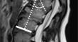
Methods: All spinal MRI scans in MPS IH patients were retrospectively reviewed. The presence, spinal location, and angulation of the gibbus deformities were collected. The angles between the superior endplate of the superior normal vertebral body and the inferior endplate of the inferior normal vertebral body were measured.
Results: 24 of 47 patients (51%) were found to have cervico-thoracic deformity on their cervical MRI scans, and 19 of those 24 (79%) patients were found to have progressive cervico-thoracic deformity with average change of angle of 17.1 degrees [range 3.9, 62.8] over 5.3 years. 7 of 8 patients who had thoraco-lumbar MRI were found to have thoraco-lumbar deformity, and 4 of those 7 patients (57%) were found to have progressive thoraco-lumbar deformity with the average increase angle of 16.7 degrees [range 3.3, 47.1] over an average of 4.1 years.
Conclusion: We found out that baseline spinal measurement cannot reliably predict the progression as multiple patients with normal alignment eventually developed severe deformity, whereases patients with severe deformity did not progress to require surgical intervention.
期刊介绍:
Neuroradiology aims to provide state-of-the-art medical and scientific information in the fields of Neuroradiology, Neurosciences, Neurology, Psychiatry, Neurosurgery, and related medical specialities. Neuroradiology as the official Journal of the European Society of Neuroradiology receives submissions from all parts of the world and publishes peer-reviewed original research, comprehensive reviews, educational papers, opinion papers, and short reports on exceptional clinical observations and new technical developments in the field of Neuroimaging and Neurointervention. The journal has subsections for Diagnostic and Interventional Neuroradiology, Advanced Neuroimaging, Paediatric Neuroradiology, Head-Neck-ENT Radiology, Spine Neuroradiology, and for submissions from Japan. Neuroradiology aims to provide new knowledge about and insights into the function and pathology of the human nervous system that may help to better diagnose and treat nervous system diseases. Neuroradiology is a member of the Committee on Publication Ethics (COPE) and follows the COPE core practices. Neuroradiology prefers articles that are free of bias, self-critical regarding limitations, transparent and clear in describing study participants, methods, and statistics, and short in presenting results. Before peer-review all submissions are automatically checked by iThenticate to assess for potential overlap in prior publication.


 分享
分享
 求助内容:
求助内容: 应助结果提醒方式:
 扫码关注我们
