Aaron Schroeder , Kai Ostendorf , Kathrin Bäumler , Domenico Mastrodicasa , Veit Sandfort , Dominik Fleischmann , Bernhard Preim , Gabriel Mistelbauer
{"title":"主动脉夹层解剖和血流动力学高级可视化","authors":"Aaron Schroeder , Kai Ostendorf , Kathrin Bäumler , Domenico Mastrodicasa , Veit Sandfort , Dominik Fleischmann , Bernhard Preim , Gabriel Mistelbauer","doi":"10.1016/j.cag.2024.104060","DOIUrl":null,"url":null,"abstract":"<div><p>Aortic dissection is a life-threatening cardiovascular disease constituted by the delamination of the aortic wall. Due to the weakened structure of the false lumen, the aorta often dilates over time, which can – after certain diameter thresholds are reached – increase the risk of fatal aortic rupture. The identification of patients with a high risk of late adverse events is an ongoing clinical challenge, further complicated by the complex dissection anatomy and the wide variety among patients. Moreover, patient-specific risk stratification depends not only on morphological, but also on hemodynamic factors, which can be derived from computer simulations or 4D flow magnetic resonance imaging (MRI). However, comprehensible visualizations that depict the complex anatomical and functional information in a single view are yet to be developed. These visualization tools will assist clinical research and decision-making by facilitating a comprehensive understanding of the aortic state. For that purpose, we identified several visualization tasks and requirements in close collaboration with cardiovascular imaging scientists and radiologists. We displayed true and false lumen hemodynamics using pathlines as well as surface hemodynamics on the dissection flap and the inner vessel wall. Pathlines indicate antegrade and retrograde flow, blood flow through fenestrations, and branch vessel supply. Dissection-specific hemodynamic measures, such as interluminal pressure difference and flap compliance, provide further insight of the blood flow throughout the cardiac cycle. Finally, we evaluated our visualization techniques with cardiothoracic and vascular surgeons in two separate virtual sessions.</p></div>","PeriodicalId":50628,"journal":{"name":"Computers & Graphics-Uk","volume":"124 ","pages":"Article 104060"},"PeriodicalIF":2.8000,"publicationDate":"2024-11-01","publicationTypes":"Journal Article","fieldsOfStudy":null,"isOpenAccess":false,"openAccessPdf":"https://www.sciencedirect.com/science/article/pii/S009784932400195X/pdfft?md5=0ad3789d79a9874f94f737d74b5f7695&pid=1-s2.0-S009784932400195X-main.pdf","citationCount":"0","resultStr":"{\"title\":\"Advanced visualization of aortic dissection anatomy and hemodynamics\",\"authors\":\"Aaron Schroeder , Kai Ostendorf , Kathrin Bäumler , Domenico Mastrodicasa , Veit Sandfort , Dominik Fleischmann , Bernhard Preim , Gabriel Mistelbauer\",\"doi\":\"10.1016/j.cag.2024.104060\",\"DOIUrl\":null,\"url\":null,\"abstract\":\"<div><p>Aortic dissection is a life-threatening cardiovascular disease constituted by the delamination of the aortic wall. Due to the weakened structure of the false lumen, the aorta often dilates over time, which can – after certain diameter thresholds are reached – increase the risk of fatal aortic rupture. The identification of patients with a high risk of late adverse events is an ongoing clinical challenge, further complicated by the complex dissection anatomy and the wide variety among patients. Moreover, patient-specific risk stratification depends not only on morphological, but also on hemodynamic factors, which can be derived from computer simulations or 4D flow magnetic resonance imaging (MRI). However, comprehensible visualizations that depict the complex anatomical and functional information in a single view are yet to be developed. These visualization tools will assist clinical research and decision-making by facilitating a comprehensive understanding of the aortic state. For that purpose, we identified several visualization tasks and requirements in close collaboration with cardiovascular imaging scientists and radiologists. We displayed true and false lumen hemodynamics using pathlines as well as surface hemodynamics on the dissection flap and the inner vessel wall. Pathlines indicate antegrade and retrograde flow, blood flow through fenestrations, and branch vessel supply. Dissection-specific hemodynamic measures, such as interluminal pressure difference and flap compliance, provide further insight of the blood flow throughout the cardiac cycle. Finally, we evaluated our visualization techniques with cardiothoracic and vascular surgeons in two separate virtual sessions.</p></div>\",\"PeriodicalId\":50628,\"journal\":{\"name\":\"Computers & Graphics-Uk\",\"volume\":\"124 \",\"pages\":\"Article 104060\"},\"PeriodicalIF\":2.8000,\"publicationDate\":\"2024-11-01\",\"publicationTypes\":\"Journal Article\",\"fieldsOfStudy\":null,\"isOpenAccess\":false,\"openAccessPdf\":\"https://www.sciencedirect.com/science/article/pii/S009784932400195X/pdfft?md5=0ad3789d79a9874f94f737d74b5f7695&pid=1-s2.0-S009784932400195X-main.pdf\",\"citationCount\":\"0\",\"resultStr\":null,\"platform\":\"Semanticscholar\",\"paperid\":null,\"PeriodicalName\":\"Computers & Graphics-Uk\",\"FirstCategoryId\":\"94\",\"ListUrlMain\":\"https://www.sciencedirect.com/science/article/pii/S009784932400195X\",\"RegionNum\":4,\"RegionCategory\":\"计算机科学\",\"ArticlePicture\":[],\"TitleCN\":null,\"AbstractTextCN\":null,\"PMCID\":null,\"EPubDate\":\"2024/8/30 0:00:00\",\"PubModel\":\"Epub\",\"JCR\":\"Q2\",\"JCRName\":\"COMPUTER SCIENCE, SOFTWARE ENGINEERING\",\"Score\":null,\"Total\":0}","platform":"Semanticscholar","paperid":null,"PeriodicalName":"Computers & Graphics-Uk","FirstCategoryId":"94","ListUrlMain":"https://www.sciencedirect.com/science/article/pii/S009784932400195X","RegionNum":4,"RegionCategory":"计算机科学","ArticlePicture":[],"TitleCN":null,"AbstractTextCN":null,"PMCID":null,"EPubDate":"2024/8/30 0:00:00","PubModel":"Epub","JCR":"Q2","JCRName":"COMPUTER SCIENCE, SOFTWARE ENGINEERING","Score":null,"Total":0}
Advanced visualization of aortic dissection anatomy and hemodynamics
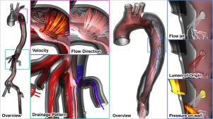
Aortic dissection is a life-threatening cardiovascular disease constituted by the delamination of the aortic wall. Due to the weakened structure of the false lumen, the aorta often dilates over time, which can – after certain diameter thresholds are reached – increase the risk of fatal aortic rupture. The identification of patients with a high risk of late adverse events is an ongoing clinical challenge, further complicated by the complex dissection anatomy and the wide variety among patients. Moreover, patient-specific risk stratification depends not only on morphological, but also on hemodynamic factors, which can be derived from computer simulations or 4D flow magnetic resonance imaging (MRI). However, comprehensible visualizations that depict the complex anatomical and functional information in a single view are yet to be developed. These visualization tools will assist clinical research and decision-making by facilitating a comprehensive understanding of the aortic state. For that purpose, we identified several visualization tasks and requirements in close collaboration with cardiovascular imaging scientists and radiologists. We displayed true and false lumen hemodynamics using pathlines as well as surface hemodynamics on the dissection flap and the inner vessel wall. Pathlines indicate antegrade and retrograde flow, blood flow through fenestrations, and branch vessel supply. Dissection-specific hemodynamic measures, such as interluminal pressure difference and flap compliance, provide further insight of the blood flow throughout the cardiac cycle. Finally, we evaluated our visualization techniques with cardiothoracic and vascular surgeons in two separate virtual sessions.
期刊介绍:
Computers & Graphics is dedicated to disseminate information on research and applications of computer graphics (CG) techniques. The journal encourages articles on:
1. Research and applications of interactive computer graphics. We are particularly interested in novel interaction techniques and applications of CG to problem domains.
2. State-of-the-art papers on late-breaking, cutting-edge research on CG.
3. Information on innovative uses of graphics principles and technologies.
4. Tutorial papers on both teaching CG principles and innovative uses of CG in education.


 分享
分享
 求助内容:
求助内容: 应助结果提醒方式:
 扫码关注我们
