Matteo Figini, Hongxiang Lin, Felice D'Arco, Godwin Ogbole, Maria Camilla Rossi-Espagnet, Olalekan Ibukun Oyinloye, Joseph Yaria, Donald Amasike Nzeh, Mojisola Omolola Atalabi, David W Carmichael, Judith Helen Cross, Ikeoluwa Lagunju, Delmiro Fernandez-Reyes, Daniel C Alexander
{"title":"利用图像质量转移对低场磁共振成像中癫痫病灶可视化增强的评估:发展中国家临床应用潜力的初步调查。","authors":"Matteo Figini, Hongxiang Lin, Felice D'Arco, Godwin Ogbole, Maria Camilla Rossi-Espagnet, Olalekan Ibukun Oyinloye, Joseph Yaria, Donald Amasike Nzeh, Mojisola Omolola Atalabi, David W Carmichael, Judith Helen Cross, Ikeoluwa Lagunju, Delmiro Fernandez-Reyes, Daniel C Alexander","doi":"10.1007/s00234-024-03448-2","DOIUrl":null,"url":null,"abstract":"<p><strong>Purpose: </strong>Low-field (LF) MRI scanners are common in many Low- and middle-Income countries, but they provide images with worse spatial resolution and contrast than high-field (HF) scanners. Image Quality Transfer (IQT) is a machine learning framework to enhance images based on high-quality references that has recently adapted to LF MRI. In this study we aim to assess if it can improve lesion visualisation compared to LF MRI scans in children with epilepsy.</p><p><strong>Methods: </strong>T1-weighted, T2-weighted and FLAIR were acquired from 12 patients (5 to 18 years old, 7 males) with clinical diagnosis of intractable epilepsy on a 0.36T (LF) and a 1.5T scanner (HF). LF images were enhanced with IQT. Seven radiologists blindly evaluated the differentiation between normal grey matter (GM) and white matter (WM) and the extension and definition of epileptogenic lesions in LF, HF and IQT-enhanced images.</p><p><strong>Results: </strong>When images were evaluated independently, GM-WM differentiation scores of IQT outputs were 26% higher, 17% higher and 12% lower than LF for T1, T2 and FLAIR. Lesion definition scores were 8-34% lower than LF, but became 3% higher than LF for FLAIR and T1 when images were seen side by side. Radiologists with expertise at HF scored IQT images higher than those with expertise at LF.</p><p><strong>Conclusion: </strong>IQT generally improved the image quality assessments. Evaluation of pathology on IQT-enhanced images was affected by familiarity with HF/IQT image appearance. These preliminary results show that IQT could have an important impact on neuroradiology practice where HF MRI is not available.</p>","PeriodicalId":19422,"journal":{"name":"Neuroradiology","volume":" ","pages":"2243-2252"},"PeriodicalIF":2.6000,"publicationDate":"2024-12-01","publicationTypes":"Journal Article","fieldsOfStudy":null,"isOpenAccess":false,"openAccessPdf":"https://www.ncbi.nlm.nih.gov/pmc/articles/PMC11611997/pdf/","citationCount":"0","resultStr":"{\"title\":\"Evaluation of epilepsy lesion visualisation enhancement in low-field MRI using image quality transfer: a preliminary investigation of clinical potential for applications in developing countries.\",\"authors\":\"Matteo Figini, Hongxiang Lin, Felice D'Arco, Godwin Ogbole, Maria Camilla Rossi-Espagnet, Olalekan Ibukun Oyinloye, Joseph Yaria, Donald Amasike Nzeh, Mojisola Omolola Atalabi, David W Carmichael, Judith Helen Cross, Ikeoluwa Lagunju, Delmiro Fernandez-Reyes, Daniel C Alexander\",\"doi\":\"10.1007/s00234-024-03448-2\",\"DOIUrl\":null,\"url\":null,\"abstract\":\"<p><strong>Purpose: </strong>Low-field (LF) MRI scanners are common in many Low- and middle-Income countries, but they provide images with worse spatial resolution and contrast than high-field (HF) scanners. Image Quality Transfer (IQT) is a machine learning framework to enhance images based on high-quality references that has recently adapted to LF MRI. In this study we aim to assess if it can improve lesion visualisation compared to LF MRI scans in children with epilepsy.</p><p><strong>Methods: </strong>T1-weighted, T2-weighted and FLAIR were acquired from 12 patients (5 to 18 years old, 7 males) with clinical diagnosis of intractable epilepsy on a 0.36T (LF) and a 1.5T scanner (HF). LF images were enhanced with IQT. Seven radiologists blindly evaluated the differentiation between normal grey matter (GM) and white matter (WM) and the extension and definition of epileptogenic lesions in LF, HF and IQT-enhanced images.</p><p><strong>Results: </strong>When images were evaluated independently, GM-WM differentiation scores of IQT outputs were 26% higher, 17% higher and 12% lower than LF for T1, T2 and FLAIR. Lesion definition scores were 8-34% lower than LF, but became 3% higher than LF for FLAIR and T1 when images were seen side by side. Radiologists with expertise at HF scored IQT images higher than those with expertise at LF.</p><p><strong>Conclusion: </strong>IQT generally improved the image quality assessments. Evaluation of pathology on IQT-enhanced images was affected by familiarity with HF/IQT image appearance. These preliminary results show that IQT could have an important impact on neuroradiology practice where HF MRI is not available.</p>\",\"PeriodicalId\":19422,\"journal\":{\"name\":\"Neuroradiology\",\"volume\":\" \",\"pages\":\"2243-2252\"},\"PeriodicalIF\":2.6000,\"publicationDate\":\"2024-12-01\",\"publicationTypes\":\"Journal Article\",\"fieldsOfStudy\":null,\"isOpenAccess\":false,\"openAccessPdf\":\"https://www.ncbi.nlm.nih.gov/pmc/articles/PMC11611997/pdf/\",\"citationCount\":\"0\",\"resultStr\":null,\"platform\":\"Semanticscholar\",\"paperid\":null,\"PeriodicalName\":\"Neuroradiology\",\"FirstCategoryId\":\"3\",\"ListUrlMain\":\"https://doi.org/10.1007/s00234-024-03448-2\",\"RegionNum\":3,\"RegionCategory\":\"医学\",\"ArticlePicture\":[],\"TitleCN\":null,\"AbstractTextCN\":null,\"PMCID\":null,\"EPubDate\":\"2024/9/6 0:00:00\",\"PubModel\":\"Epub\",\"JCR\":\"Q2\",\"JCRName\":\"CLINICAL NEUROLOGY\",\"Score\":null,\"Total\":0}","platform":"Semanticscholar","paperid":null,"PeriodicalName":"Neuroradiology","FirstCategoryId":"3","ListUrlMain":"https://doi.org/10.1007/s00234-024-03448-2","RegionNum":3,"RegionCategory":"医学","ArticlePicture":[],"TitleCN":null,"AbstractTextCN":null,"PMCID":null,"EPubDate":"2024/9/6 0:00:00","PubModel":"Epub","JCR":"Q2","JCRName":"CLINICAL NEUROLOGY","Score":null,"Total":0}
Evaluation of epilepsy lesion visualisation enhancement in low-field MRI using image quality transfer: a preliminary investigation of clinical potential for applications in developing countries.
Purpose: Low-field (LF) MRI scanners are common in many Low- and middle-Income countries, but they provide images with worse spatial resolution and contrast than high-field (HF) scanners. Image Quality Transfer (IQT) is a machine learning framework to enhance images based on high-quality references that has recently adapted to LF MRI. In this study we aim to assess if it can improve lesion visualisation compared to LF MRI scans in children with epilepsy.
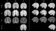
Methods: T1-weighted, T2-weighted and FLAIR were acquired from 12 patients (5 to 18 years old, 7 males) with clinical diagnosis of intractable epilepsy on a 0.36T (LF) and a 1.5T scanner (HF). LF images were enhanced with IQT. Seven radiologists blindly evaluated the differentiation between normal grey matter (GM) and white matter (WM) and the extension and definition of epileptogenic lesions in LF, HF and IQT-enhanced images.
Results: When images were evaluated independently, GM-WM differentiation scores of IQT outputs were 26% higher, 17% higher and 12% lower than LF for T1, T2 and FLAIR. Lesion definition scores were 8-34% lower than LF, but became 3% higher than LF for FLAIR and T1 when images were seen side by side. Radiologists with expertise at HF scored IQT images higher than those with expertise at LF.
Conclusion: IQT generally improved the image quality assessments. Evaluation of pathology on IQT-enhanced images was affected by familiarity with HF/IQT image appearance. These preliminary results show that IQT could have an important impact on neuroradiology practice where HF MRI is not available.
期刊介绍:
Neuroradiology aims to provide state-of-the-art medical and scientific information in the fields of Neuroradiology, Neurosciences, Neurology, Psychiatry, Neurosurgery, and related medical specialities. Neuroradiology as the official Journal of the European Society of Neuroradiology receives submissions from all parts of the world and publishes peer-reviewed original research, comprehensive reviews, educational papers, opinion papers, and short reports on exceptional clinical observations and new technical developments in the field of Neuroimaging and Neurointervention. The journal has subsections for Diagnostic and Interventional Neuroradiology, Advanced Neuroimaging, Paediatric Neuroradiology, Head-Neck-ENT Radiology, Spine Neuroradiology, and for submissions from Japan. Neuroradiology aims to provide new knowledge about and insights into the function and pathology of the human nervous system that may help to better diagnose and treat nervous system diseases. Neuroradiology is a member of the Committee on Publication Ethics (COPE) and follows the COPE core practices. Neuroradiology prefers articles that are free of bias, self-critical regarding limitations, transparent and clear in describing study participants, methods, and statistics, and short in presenting results. Before peer-review all submissions are automatically checked by iThenticate to assess for potential overlap in prior publication.


 分享
分享
 求助内容:
求助内容: 应助结果提醒方式:
 扫码关注我们
