Kalli K. Stephens, Ryan M. Finnerty, DeAna G. Grant, Sarayut Winuthayanon, Patricia A. Martin-DeLeon, Wipawee Winuthayanon
{"title":"植入前胚胎发育过程中小鼠输卵管细胞外囊泡的蛋白质组分析和活体可视化。","authors":"Kalli K. Stephens, Ryan M. Finnerty, DeAna G. Grant, Sarayut Winuthayanon, Patricia A. Martin-DeLeon, Wipawee Winuthayanon","doi":"10.1096/fj.202400041RR","DOIUrl":null,"url":null,"abstract":"<p>Pre-implantation embryonic development occurs in the oviduct during the first few days of pregnancy. The presence of oviductal extracellular vesicles (oEVs, also called oviductosomes) is crucial for pre-implantation embryonic development in vivo as oEVs often contain molecular transmitters such as proteins. Therefore, evaluating oEV cargo during early pregnancy could provide insights into factors required for proper early embryonic development that are missing in the current in vitro embryo culture setting. In this study, we isolated oEVs from the oviductal fluid at estrus and different stages of early embryonic development. The 2306–3066 proteins in oEVs identified at the different time points revealed 58–60 common EV markers identified in exosome databases. Oviductal extracellular vesicle proteins from pregnant samples significantly differed from those in non-pregnant samples. In addition, superovulation changes the protein contents in oEVs compared to natural ovulation at estrus. Importantly, we have identified that embryo-protectant proteins such as high-mobility protein group B1 and serine (or cysteine) peptidase inhibitor were only enriched in the presence of embryos. We also visualized the physical interaction of EVs and the zona pellucida of 4- to 8-cell stage embryos using transmission electron microscopy as well as in vivo live imaging of epithelial cell-derived GFP-tagged CD9 mouse model. All protein data in this study are readily available to the scientific community in a searchable format at https://genes.winuthayanon.com/winuthayanon/oviduct_ev_proteins/. In conclusion, we identified oEVs proteins that could be tested to determine whether they can improve embryonic developmental outcomes in vivo and in vitro setting.</p>","PeriodicalId":50455,"journal":{"name":"The FASEB Journal","volume":"38 17","pages":""},"PeriodicalIF":4.2000,"publicationDate":"2024-09-06","publicationTypes":"Journal Article","fieldsOfStudy":null,"isOpenAccess":false,"openAccessPdf":"","citationCount":"0","resultStr":"{\"title\":\"Proteomic analysis and in vivo visualization of extracellular vesicles from mouse oviducts during pre-implantation embryo development\",\"authors\":\"Kalli K. Stephens, Ryan M. Finnerty, DeAna G. Grant, Sarayut Winuthayanon, Patricia A. Martin-DeLeon, Wipawee Winuthayanon\",\"doi\":\"10.1096/fj.202400041RR\",\"DOIUrl\":null,\"url\":null,\"abstract\":\"<p>Pre-implantation embryonic development occurs in the oviduct during the first few days of pregnancy. The presence of oviductal extracellular vesicles (oEVs, also called oviductosomes) is crucial for pre-implantation embryonic development in vivo as oEVs often contain molecular transmitters such as proteins. Therefore, evaluating oEV cargo during early pregnancy could provide insights into factors required for proper early embryonic development that are missing in the current in vitro embryo culture setting. In this study, we isolated oEVs from the oviductal fluid at estrus and different stages of early embryonic development. The 2306–3066 proteins in oEVs identified at the different time points revealed 58–60 common EV markers identified in exosome databases. Oviductal extracellular vesicle proteins from pregnant samples significantly differed from those in non-pregnant samples. In addition, superovulation changes the protein contents in oEVs compared to natural ovulation at estrus. Importantly, we have identified that embryo-protectant proteins such as high-mobility protein group B1 and serine (or cysteine) peptidase inhibitor were only enriched in the presence of embryos. We also visualized the physical interaction of EVs and the zona pellucida of 4- to 8-cell stage embryos using transmission electron microscopy as well as in vivo live imaging of epithelial cell-derived GFP-tagged CD9 mouse model. All protein data in this study are readily available to the scientific community in a searchable format at https://genes.winuthayanon.com/winuthayanon/oviduct_ev_proteins/. In conclusion, we identified oEVs proteins that could be tested to determine whether they can improve embryonic developmental outcomes in vivo and in vitro setting.</p>\",\"PeriodicalId\":50455,\"journal\":{\"name\":\"The FASEB Journal\",\"volume\":\"38 17\",\"pages\":\"\"},\"PeriodicalIF\":4.2000,\"publicationDate\":\"2024-09-06\",\"publicationTypes\":\"Journal Article\",\"fieldsOfStudy\":null,\"isOpenAccess\":false,\"openAccessPdf\":\"\",\"citationCount\":\"0\",\"resultStr\":null,\"platform\":\"Semanticscholar\",\"paperid\":null,\"PeriodicalName\":\"The FASEB Journal\",\"FirstCategoryId\":\"99\",\"ListUrlMain\":\"https://faseb.onlinelibrary.wiley.com/doi/10.1096/fj.202400041RR\",\"RegionNum\":2,\"RegionCategory\":\"生物学\",\"ArticlePicture\":[],\"TitleCN\":null,\"AbstractTextCN\":null,\"PMCID\":null,\"EPubDate\":\"\",\"PubModel\":\"\",\"JCR\":\"Q2\",\"JCRName\":\"BIOCHEMISTRY & MOLECULAR BIOLOGY\",\"Score\":null,\"Total\":0}","platform":"Semanticscholar","paperid":null,"PeriodicalName":"The FASEB Journal","FirstCategoryId":"99","ListUrlMain":"https://faseb.onlinelibrary.wiley.com/doi/10.1096/fj.202400041RR","RegionNum":2,"RegionCategory":"生物学","ArticlePicture":[],"TitleCN":null,"AbstractTextCN":null,"PMCID":null,"EPubDate":"","PubModel":"","JCR":"Q2","JCRName":"BIOCHEMISTRY & MOLECULAR BIOLOGY","Score":null,"Total":0}
Proteomic analysis and in vivo visualization of extracellular vesicles from mouse oviducts during pre-implantation embryo development
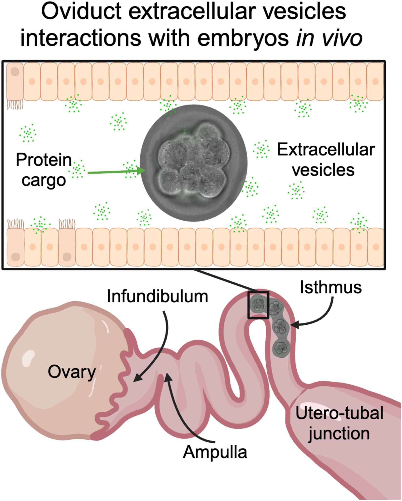
Pre-implantation embryonic development occurs in the oviduct during the first few days of pregnancy. The presence of oviductal extracellular vesicles (oEVs, also called oviductosomes) is crucial for pre-implantation embryonic development in vivo as oEVs often contain molecular transmitters such as proteins. Therefore, evaluating oEV cargo during early pregnancy could provide insights into factors required for proper early embryonic development that are missing in the current in vitro embryo culture setting. In this study, we isolated oEVs from the oviductal fluid at estrus and different stages of early embryonic development. The 2306–3066 proteins in oEVs identified at the different time points revealed 58–60 common EV markers identified in exosome databases. Oviductal extracellular vesicle proteins from pregnant samples significantly differed from those in non-pregnant samples. In addition, superovulation changes the protein contents in oEVs compared to natural ovulation at estrus. Importantly, we have identified that embryo-protectant proteins such as high-mobility protein group B1 and serine (or cysteine) peptidase inhibitor were only enriched in the presence of embryos. We also visualized the physical interaction of EVs and the zona pellucida of 4- to 8-cell stage embryos using transmission electron microscopy as well as in vivo live imaging of epithelial cell-derived GFP-tagged CD9 mouse model. All protein data in this study are readily available to the scientific community in a searchable format at https://genes.winuthayanon.com/winuthayanon/oviduct_ev_proteins/. In conclusion, we identified oEVs proteins that could be tested to determine whether they can improve embryonic developmental outcomes in vivo and in vitro setting.
期刊介绍:
The FASEB Journal publishes international, transdisciplinary research covering all fields of biology at every level of organization: atomic, molecular, cell, tissue, organ, organismic and population. While the journal strives to include research that cuts across the biological sciences, it also considers submissions that lie within one field, but may have implications for other fields as well. The journal seeks to publish basic and translational research, but also welcomes reports of pre-clinical and early clinical research. In addition to research, review, and hypothesis submissions, The FASEB Journal also seeks perspectives, commentaries, book reviews, and similar content related to the life sciences in its Up Front section.


 分享
分享
 求助内容:
求助内容: 应助结果提醒方式:
 扫码关注我们
