Laura Mawdsley, Rasa Eskandari, Farah Kamar, Ajay Rajaram, Lawrence C. M. Yip, Naomi Abayomi, Stephanie Milkovich, Jeffrey J. L. Carson, Keith St. Lawrence, Christopher G. Ellis, Mamadou Diop
{"title":"体内光学评估大脑和骨骼肌微血管对苯肾上腺素的反应","authors":"Laura Mawdsley, Rasa Eskandari, Farah Kamar, Ajay Rajaram, Lawrence C. M. Yip, Naomi Abayomi, Stephanie Milkovich, Jeffrey J. L. Carson, Keith St. Lawrence, Christopher G. Ellis, Mamadou Diop","doi":"10.1096/fba.2024-00063","DOIUrl":null,"url":null,"abstract":"<p>This study aimed to investigate the simultaneous response of the cerebral and skeletal muscle microvasculature to the same phenylephrine (PE) boluses. A hybrid optical system that combines hyperspectral near-infrared spectroscopy (hs-NIRS) and diffuse correlation spectroscopy (DCS) was used to monitor changes in tissue oxygenation and perfusion. Data were collected from the head and hind limb of seven male Sprague–Dawley rats while administering intravenous (IV) injections of PE or saline to all animals. The response to saline was used as a control. Skeletal muscle oxygenation decreased significantly after PE injection, while a statistically underpowered decrease in perfusion was observed, followed by an increase beyond baseline. Vascular conductance also decreased in the muscle reflecting the drug's vasoconstrictive effects. Tissue oxygenation and perfusion increased in the brain in response to PE. Initially, there was a sharp increase in cerebral perfusion but no changes in cerebral vascular conductance. Subsequently, cerebral flow and vascular conductance decreased significantly below baseline, likely reflecting autoregulatory mechanisms to manage the excess flow. Further, fitting an exponential function to the secondary decrease in cerebral perfusion and increase in muscular blood flow revealed a quicker kinetic response in the brain to adjust blood flow. In the skeletal muscle, PE caused a transient decrease in blood volume due to vasoconstriction, which resulted in an overall decrease in hemoglobin content and tissue oxygen saturation. Since PE does not directly affect cerebral vessels, this peripheral vasoconstriction shunted blood into the brain, resulting in an initial increase in oxygenated hemoglobin and oxygen saturation.</p>","PeriodicalId":12093,"journal":{"name":"FASEB bioAdvances","volume":"6 9","pages":"390-399"},"PeriodicalIF":2.4000,"publicationDate":"2024-08-21","publicationTypes":"Journal Article","fieldsOfStudy":null,"isOpenAccess":false,"openAccessPdf":"https://onlinelibrary.wiley.com/doi/epdf/10.1096/fba.2024-00063","citationCount":"0","resultStr":"{\"title\":\"In vivo optical assessment of cerebral and skeletal muscle microvascular response to phenylephrine\",\"authors\":\"Laura Mawdsley, Rasa Eskandari, Farah Kamar, Ajay Rajaram, Lawrence C. M. Yip, Naomi Abayomi, Stephanie Milkovich, Jeffrey J. L. Carson, Keith St. Lawrence, Christopher G. Ellis, Mamadou Diop\",\"doi\":\"10.1096/fba.2024-00063\",\"DOIUrl\":null,\"url\":null,\"abstract\":\"<p>This study aimed to investigate the simultaneous response of the cerebral and skeletal muscle microvasculature to the same phenylephrine (PE) boluses. A hybrid optical system that combines hyperspectral near-infrared spectroscopy (hs-NIRS) and diffuse correlation spectroscopy (DCS) was used to monitor changes in tissue oxygenation and perfusion. Data were collected from the head and hind limb of seven male Sprague–Dawley rats while administering intravenous (IV) injections of PE or saline to all animals. The response to saline was used as a control. Skeletal muscle oxygenation decreased significantly after PE injection, while a statistically underpowered decrease in perfusion was observed, followed by an increase beyond baseline. Vascular conductance also decreased in the muscle reflecting the drug's vasoconstrictive effects. Tissue oxygenation and perfusion increased in the brain in response to PE. Initially, there was a sharp increase in cerebral perfusion but no changes in cerebral vascular conductance. Subsequently, cerebral flow and vascular conductance decreased significantly below baseline, likely reflecting autoregulatory mechanisms to manage the excess flow. Further, fitting an exponential function to the secondary decrease in cerebral perfusion and increase in muscular blood flow revealed a quicker kinetic response in the brain to adjust blood flow. In the skeletal muscle, PE caused a transient decrease in blood volume due to vasoconstriction, which resulted in an overall decrease in hemoglobin content and tissue oxygen saturation. Since PE does not directly affect cerebral vessels, this peripheral vasoconstriction shunted blood into the brain, resulting in an initial increase in oxygenated hemoglobin and oxygen saturation.</p>\",\"PeriodicalId\":12093,\"journal\":{\"name\":\"FASEB bioAdvances\",\"volume\":\"6 9\",\"pages\":\"390-399\"},\"PeriodicalIF\":2.4000,\"publicationDate\":\"2024-08-21\",\"publicationTypes\":\"Journal Article\",\"fieldsOfStudy\":null,\"isOpenAccess\":false,\"openAccessPdf\":\"https://onlinelibrary.wiley.com/doi/epdf/10.1096/fba.2024-00063\",\"citationCount\":\"0\",\"resultStr\":null,\"platform\":\"Semanticscholar\",\"paperid\":null,\"PeriodicalName\":\"FASEB bioAdvances\",\"FirstCategoryId\":\"1085\",\"ListUrlMain\":\"https://faseb.onlinelibrary.wiley.com/doi/10.1096/fba.2024-00063\",\"RegionNum\":0,\"RegionCategory\":null,\"ArticlePicture\":[],\"TitleCN\":null,\"AbstractTextCN\":null,\"PMCID\":null,\"EPubDate\":\"\",\"PubModel\":\"\",\"JCR\":\"Q3\",\"JCRName\":\"BIOCHEMISTRY & MOLECULAR BIOLOGY\",\"Score\":null,\"Total\":0}","platform":"Semanticscholar","paperid":null,"PeriodicalName":"FASEB bioAdvances","FirstCategoryId":"1085","ListUrlMain":"https://faseb.onlinelibrary.wiley.com/doi/10.1096/fba.2024-00063","RegionNum":0,"RegionCategory":null,"ArticlePicture":[],"TitleCN":null,"AbstractTextCN":null,"PMCID":null,"EPubDate":"","PubModel":"","JCR":"Q3","JCRName":"BIOCHEMISTRY & MOLECULAR BIOLOGY","Score":null,"Total":0}
引用次数: 0
摘要
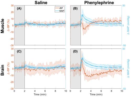
本研究旨在探讨大脑和骨骼肌微血管对相同的苯肾上腺素(PE)栓剂的同时反应。研究使用了一种结合了高光谱近红外光谱(hs-NIRS)和弥散相关光谱(DCS)的混合光学系统来监测组织氧合和灌注的变化。在对所有动物静脉注射 PE 或生理盐水的同时,从七只雄性 Sprague-Dawley 大鼠的头部和后肢收集数据。对生理盐水的反应作为对照。注射 PE 后,骨骼肌氧饱和度明显下降,同时观察到灌注量出现统计学意义上的下降,随后又超过基线上升。肌肉中的血管传导性也有所下降,这反映了药物的血管收缩效应。脑组织含氧量和灌注量在 PE 作用下有所增加。最初,脑灌注急剧增加,但脑血管传导没有变化。随后,脑血流和血管传导显著下降,低于基线,这可能反映了管理过量血流的自动调节机制。此外,用指数函数拟合脑灌注的继发性减少和肌肉血流量的增加,发现大脑对调整血流量有较快的动力学反应。在骨骼肌中,由于血管收缩,PE 会导致血容量短暂减少,从而导致血红蛋白含量和组织氧饱和度整体下降。由于 PE 并不直接影响脑血管,这种外周血管收缩会将血液分流到大脑,从而导致氧合血红蛋白和氧饱和度的最初增加。
In vivo optical assessment of cerebral and skeletal muscle microvascular response to phenylephrine
This study aimed to investigate the simultaneous response of the cerebral and skeletal muscle microvasculature to the same phenylephrine (PE) boluses. A hybrid optical system that combines hyperspectral near-infrared spectroscopy (hs-NIRS) and diffuse correlation spectroscopy (DCS) was used to monitor changes in tissue oxygenation and perfusion. Data were collected from the head and hind limb of seven male Sprague–Dawley rats while administering intravenous (IV) injections of PE or saline to all animals. The response to saline was used as a control. Skeletal muscle oxygenation decreased significantly after PE injection, while a statistically underpowered decrease in perfusion was observed, followed by an increase beyond baseline. Vascular conductance also decreased in the muscle reflecting the drug's vasoconstrictive effects. Tissue oxygenation and perfusion increased in the brain in response to PE. Initially, there was a sharp increase in cerebral perfusion but no changes in cerebral vascular conductance. Subsequently, cerebral flow and vascular conductance decreased significantly below baseline, likely reflecting autoregulatory mechanisms to manage the excess flow. Further, fitting an exponential function to the secondary decrease in cerebral perfusion and increase in muscular blood flow revealed a quicker kinetic response in the brain to adjust blood flow. In the skeletal muscle, PE caused a transient decrease in blood volume due to vasoconstriction, which resulted in an overall decrease in hemoglobin content and tissue oxygen saturation. Since PE does not directly affect cerebral vessels, this peripheral vasoconstriction shunted blood into the brain, resulting in an initial increase in oxygenated hemoglobin and oxygen saturation.


 分享
分享
 求助内容:
求助内容: 应助结果提醒方式:
 扫码关注我们
