Lisa Klaassen, Myriam G Jaarsma-Coes, Marina Marinkovic, Gregorius P M Luyten, Coen R N Rasch, Teresa A Ferreira, Jan-Willem M Beenakker
{"title":"葡萄膜黑色素瘤的定量灌注加权磁共振成像","authors":"Lisa Klaassen, Myriam G Jaarsma-Coes, Marina Marinkovic, Gregorius P M Luyten, Coen R N Rasch, Teresa A Ferreira, Jan-Willem M Beenakker","doi":"10.1167/iovs.65.11.17","DOIUrl":null,"url":null,"abstract":"<p><strong>Purpose: </strong>Perfusion-weighted imaging (PWI; magnetic resonance imaging [MRI]) has been shown to provide valuable biological tumor information in uveal melanoma (UM). Clinically used semiquantitative methods do not account for tumor pigmentation and eye movement. We hypothesize that a quantitative PWI method that incorporates these, provides a more accurate description of tumor perfusion than the current clinical method. The aim of this study was to test this in patients with UM before and after radiotherapy.</p><p><strong>Methods: </strong>Perfusion-weighted 3T MRIs were retrospectively analyzed in 47 patients with UM before and after radiotherapy. Tofts pharmacokinetic modeling was performed to determine vascular permeability (Ktrans), extracellular extravascular space (ve), and reflux rate (kep). These were compared with semiquantitative clinical parameters including peak intensity and outflow percentage.</p><p><strong>Results: </strong>The effect of tumor pigmentation on peak intensity and outflow percentage was statistically significant (P < 0.01) and relative peak intensity was significantly different between melanotic and amelanotic tumors (1.5 vs. 1.9, P < 0.01). Before radiotherapy, median tumor Ktrans was 0.63 min-1 (range = 0.06-1.42 min-1), median ve was 0.23 (range = 0.09-0.63), and median kep was 2.3 min-1 (range = 0.6-5.0 min-1). After radiotherapy, 85% showed a decrease in Ktrans and kep (P < 0.01). Changes in tumor pigmentation before and after radiotherapy were small and not significant (median increase in T1 of 33 ms, P = 0.55).</p><p><strong>Conclusions: </strong>Quantitative PWI parameters decreased significantly after radiotherapy and can therefore can serve as an early biomarker for treatment response assessment. However, due to the nonsignificant changes in tumor pigmentation before and after radiotherapy, the current semiquantitative method appears to be sufficiently sensitive for detection of changes in tumor perfusion.</p>","PeriodicalId":14620,"journal":{"name":"Investigative ophthalmology & visual science","volume":"65 11","pages":"17"},"PeriodicalIF":5.5000,"publicationDate":"2024-09-03","publicationTypes":"Journal Article","fieldsOfStudy":null,"isOpenAccess":false,"openAccessPdf":"https://www.ncbi.nlm.nih.gov/pmc/articles/PMC11385876/pdf/","citationCount":"0","resultStr":"{\"title\":\"Quantitative Perfusion-Weighted Magnetic Resonance Imaging in Uveal Melanoma.\",\"authors\":\"Lisa Klaassen, Myriam G Jaarsma-Coes, Marina Marinkovic, Gregorius P M Luyten, Coen R N Rasch, Teresa A Ferreira, Jan-Willem M Beenakker\",\"doi\":\"10.1167/iovs.65.11.17\",\"DOIUrl\":null,\"url\":null,\"abstract\":\"<p><strong>Purpose: </strong>Perfusion-weighted imaging (PWI; magnetic resonance imaging [MRI]) has been shown to provide valuable biological tumor information in uveal melanoma (UM). Clinically used semiquantitative methods do not account for tumor pigmentation and eye movement. We hypothesize that a quantitative PWI method that incorporates these, provides a more accurate description of tumor perfusion than the current clinical method. The aim of this study was to test this in patients with UM before and after radiotherapy.</p><p><strong>Methods: </strong>Perfusion-weighted 3T MRIs were retrospectively analyzed in 47 patients with UM before and after radiotherapy. Tofts pharmacokinetic modeling was performed to determine vascular permeability (Ktrans), extracellular extravascular space (ve), and reflux rate (kep). These were compared with semiquantitative clinical parameters including peak intensity and outflow percentage.</p><p><strong>Results: </strong>The effect of tumor pigmentation on peak intensity and outflow percentage was statistically significant (P < 0.01) and relative peak intensity was significantly different between melanotic and amelanotic tumors (1.5 vs. 1.9, P < 0.01). Before radiotherapy, median tumor Ktrans was 0.63 min-1 (range = 0.06-1.42 min-1), median ve was 0.23 (range = 0.09-0.63), and median kep was 2.3 min-1 (range = 0.6-5.0 min-1). After radiotherapy, 85% showed a decrease in Ktrans and kep (P < 0.01). Changes in tumor pigmentation before and after radiotherapy were small and not significant (median increase in T1 of 33 ms, P = 0.55).</p><p><strong>Conclusions: </strong>Quantitative PWI parameters decreased significantly after radiotherapy and can therefore can serve as an early biomarker for treatment response assessment. However, due to the nonsignificant changes in tumor pigmentation before and after radiotherapy, the current semiquantitative method appears to be sufficiently sensitive for detection of changes in tumor perfusion.</p>\",\"PeriodicalId\":14620,\"journal\":{\"name\":\"Investigative ophthalmology & visual science\",\"volume\":\"65 11\",\"pages\":\"17\"},\"PeriodicalIF\":5.5000,\"publicationDate\":\"2024-09-03\",\"publicationTypes\":\"Journal Article\",\"fieldsOfStudy\":null,\"isOpenAccess\":false,\"openAccessPdf\":\"https://www.ncbi.nlm.nih.gov/pmc/articles/PMC11385876/pdf/\",\"citationCount\":\"0\",\"resultStr\":null,\"platform\":\"Semanticscholar\",\"paperid\":null,\"PeriodicalName\":\"Investigative ophthalmology & visual science\",\"FirstCategoryId\":\"3\",\"ListUrlMain\":\"https://doi.org/10.1167/iovs.65.11.17\",\"RegionNum\":2,\"RegionCategory\":\"医学\",\"ArticlePicture\":[],\"TitleCN\":null,\"AbstractTextCN\":null,\"PMCID\":null,\"EPubDate\":\"\",\"PubModel\":\"\",\"JCR\":\"Q1\",\"JCRName\":\"OPHTHALMOLOGY\",\"Score\":null,\"Total\":0}","platform":"Semanticscholar","paperid":null,"PeriodicalName":"Investigative ophthalmology & visual science","FirstCategoryId":"3","ListUrlMain":"https://doi.org/10.1167/iovs.65.11.17","RegionNum":2,"RegionCategory":"医学","ArticlePicture":[],"TitleCN":null,"AbstractTextCN":null,"PMCID":null,"EPubDate":"","PubModel":"","JCR":"Q1","JCRName":"OPHTHALMOLOGY","Score":null,"Total":0}
Quantitative Perfusion-Weighted Magnetic Resonance Imaging in Uveal Melanoma.
Purpose: Perfusion-weighted imaging (PWI; magnetic resonance imaging [MRI]) has been shown to provide valuable biological tumor information in uveal melanoma (UM). Clinically used semiquantitative methods do not account for tumor pigmentation and eye movement. We hypothesize that a quantitative PWI method that incorporates these, provides a more accurate description of tumor perfusion than the current clinical method. The aim of this study was to test this in patients with UM before and after radiotherapy.
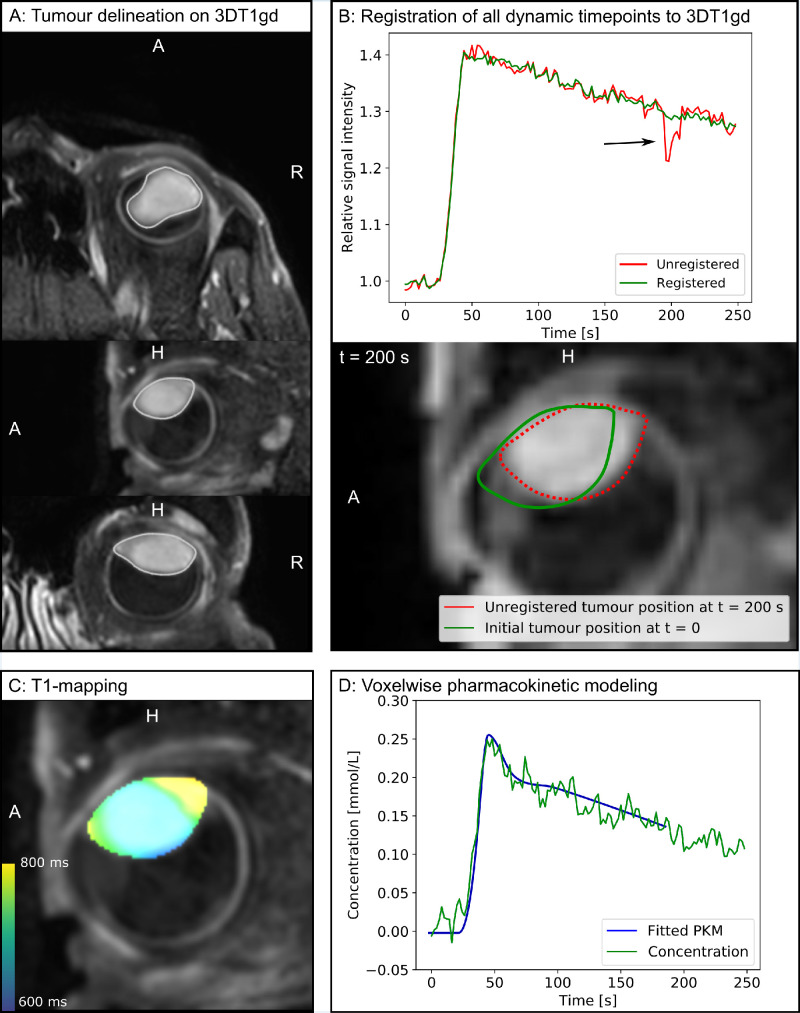
Methods: Perfusion-weighted 3T MRIs were retrospectively analyzed in 47 patients with UM before and after radiotherapy. Tofts pharmacokinetic modeling was performed to determine vascular permeability (Ktrans), extracellular extravascular space (ve), and reflux rate (kep). These were compared with semiquantitative clinical parameters including peak intensity and outflow percentage.
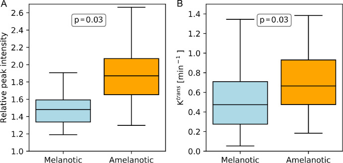
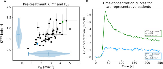
Results: The effect of tumor pigmentation on peak intensity and outflow percentage was statistically significant (P < 0.01) and relative peak intensity was significantly different between melanotic and amelanotic tumors (1.5 vs. 1.9, P < 0.01). Before radiotherapy, median tumor Ktrans was 0.63 min-1 (range = 0.06-1.42 min-1), median ve was 0.23 (range = 0.09-0.63), and median kep was 2.3 min-1 (range = 0.6-5.0 min-1). After radiotherapy, 85% showed a decrease in Ktrans and kep (P < 0.01). Changes in tumor pigmentation before and after radiotherapy were small and not significant (median increase in T1 of 33 ms, P = 0.55).
Conclusions: Quantitative PWI parameters decreased significantly after radiotherapy and can therefore can serve as an early biomarker for treatment response assessment. However, due to the nonsignificant changes in tumor pigmentation before and after radiotherapy, the current semiquantitative method appears to be sufficiently sensitive for detection of changes in tumor perfusion.
期刊介绍:
Investigative Ophthalmology & Visual Science (IOVS), published as ready online, is a peer-reviewed academic journal of the Association for Research in Vision and Ophthalmology (ARVO). IOVS features original research, mostly pertaining to clinical and laboratory ophthalmology and vision research in general.


 分享
分享

 求助内容:
求助内容: 应助结果提醒方式:
 扫码关注我们
