Jun Cao, Iain K. Ball, Benjamin Cassidy, Caroline D. Rae
{"title":"功能传导成像:大脑活动的定量绘图","authors":"Jun Cao, Iain K. Ball, Benjamin Cassidy, Caroline D. Rae","doi":"10.1007/s13246-024-01484-z","DOIUrl":null,"url":null,"abstract":"<p>Theory and modelling suggest that detection of neuronal activity may be feasible using phase sensitive MRI methods. Successful detection of neuronal activity both in vitro and in vivo has been described while others have reported negative results. Magnetic resonance electrical properties tomography may be a route by which signal changes can be identified. Here, we report successful and repeatable detection at 3 Tesla of human brain activation in response to visual and somatosensory stimuli using a functional version of tissue conductivity imaging (funCI). This detects activation in both white and grey matter with apparent tissue conductivity changes of 0.1 S/m (17–20%, depending on the tissue baseline conductivity measure) allowing visualization of complete system circuitry. The degree of activation scales with the degree of the stimulus (duration or contrast). The conductivity response functions show a distinct timecourse from that of traditional fMRI haemodynamic (BOLD or Blood Oxygenation Level Dependent) response functions, peaking within milliseconds of stimulus cessation and returning to baseline within 3–4 s. We demonstrate the utility of the funCI approach by showing robust activation of the lateral somatosensory circuitry on stimulation of an index finger, on stimulation of a big toe or of noxious (heat) stimulation of the face as well as activation of visual circuitry on visual stimulation in up to five different individuals. The sensitivity and repeatability of this approach provides further evidence that magnetic resonance imaging approaches can detect brain activation beyond changes in blood supply.</p>","PeriodicalId":48490,"journal":{"name":"Physical and Engineering Sciences in Medicine","volume":"1 1","pages":""},"PeriodicalIF":2.0000,"publicationDate":"2024-09-11","publicationTypes":"Journal Article","fieldsOfStudy":null,"isOpenAccess":false,"openAccessPdf":"","citationCount":"0","resultStr":"{\"title\":\"Functional conductivity imaging: quantitative mapping of brain activity\",\"authors\":\"Jun Cao, Iain K. Ball, Benjamin Cassidy, Caroline D. Rae\",\"doi\":\"10.1007/s13246-024-01484-z\",\"DOIUrl\":null,\"url\":null,\"abstract\":\"<p>Theory and modelling suggest that detection of neuronal activity may be feasible using phase sensitive MRI methods. Successful detection of neuronal activity both in vitro and in vivo has been described while others have reported negative results. Magnetic resonance electrical properties tomography may be a route by which signal changes can be identified. Here, we report successful and repeatable detection at 3 Tesla of human brain activation in response to visual and somatosensory stimuli using a functional version of tissue conductivity imaging (funCI). This detects activation in both white and grey matter with apparent tissue conductivity changes of 0.1 S/m (17–20%, depending on the tissue baseline conductivity measure) allowing visualization of complete system circuitry. The degree of activation scales with the degree of the stimulus (duration or contrast). The conductivity response functions show a distinct timecourse from that of traditional fMRI haemodynamic (BOLD or Blood Oxygenation Level Dependent) response functions, peaking within milliseconds of stimulus cessation and returning to baseline within 3–4 s. We demonstrate the utility of the funCI approach by showing robust activation of the lateral somatosensory circuitry on stimulation of an index finger, on stimulation of a big toe or of noxious (heat) stimulation of the face as well as activation of visual circuitry on visual stimulation in up to five different individuals. The sensitivity and repeatability of this approach provides further evidence that magnetic resonance imaging approaches can detect brain activation beyond changes in blood supply.</p>\",\"PeriodicalId\":48490,\"journal\":{\"name\":\"Physical and Engineering Sciences in Medicine\",\"volume\":\"1 1\",\"pages\":\"\"},\"PeriodicalIF\":2.0000,\"publicationDate\":\"2024-09-11\",\"publicationTypes\":\"Journal Article\",\"fieldsOfStudy\":null,\"isOpenAccess\":false,\"openAccessPdf\":\"\",\"citationCount\":\"0\",\"resultStr\":null,\"platform\":\"Semanticscholar\",\"paperid\":null,\"PeriodicalName\":\"Physical and Engineering Sciences in Medicine\",\"FirstCategoryId\":\"3\",\"ListUrlMain\":\"https://doi.org/10.1007/s13246-024-01484-z\",\"RegionNum\":4,\"RegionCategory\":\"医学\",\"ArticlePicture\":[],\"TitleCN\":null,\"AbstractTextCN\":null,\"PMCID\":null,\"EPubDate\":\"\",\"PubModel\":\"\",\"JCR\":\"Q3\",\"JCRName\":\"ENGINEERING, BIOMEDICAL\",\"Score\":null,\"Total\":0}","platform":"Semanticscholar","paperid":null,"PeriodicalName":"Physical and Engineering Sciences in Medicine","FirstCategoryId":"3","ListUrlMain":"https://doi.org/10.1007/s13246-024-01484-z","RegionNum":4,"RegionCategory":"医学","ArticlePicture":[],"TitleCN":null,"AbstractTextCN":null,"PMCID":null,"EPubDate":"","PubModel":"","JCR":"Q3","JCRName":"ENGINEERING, BIOMEDICAL","Score":null,"Total":0}
Functional conductivity imaging: quantitative mapping of brain activity
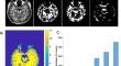
Theory and modelling suggest that detection of neuronal activity may be feasible using phase sensitive MRI methods. Successful detection of neuronal activity both in vitro and in vivo has been described while others have reported negative results. Magnetic resonance electrical properties tomography may be a route by which signal changes can be identified. Here, we report successful and repeatable detection at 3 Tesla of human brain activation in response to visual and somatosensory stimuli using a functional version of tissue conductivity imaging (funCI). This detects activation in both white and grey matter with apparent tissue conductivity changes of 0.1 S/m (17–20%, depending on the tissue baseline conductivity measure) allowing visualization of complete system circuitry. The degree of activation scales with the degree of the stimulus (duration or contrast). The conductivity response functions show a distinct timecourse from that of traditional fMRI haemodynamic (BOLD or Blood Oxygenation Level Dependent) response functions, peaking within milliseconds of stimulus cessation and returning to baseline within 3–4 s. We demonstrate the utility of the funCI approach by showing robust activation of the lateral somatosensory circuitry on stimulation of an index finger, on stimulation of a big toe or of noxious (heat) stimulation of the face as well as activation of visual circuitry on visual stimulation in up to five different individuals. The sensitivity and repeatability of this approach provides further evidence that magnetic resonance imaging approaches can detect brain activation beyond changes in blood supply.


 分享
分享
 求助内容:
求助内容: 应助结果提醒方式:
 扫码关注我们
