Frank O. F. Reilly, Ioannis Georgopoulos, Håkan Jonsson, Kevin Mani, Andrés Rodriguez-Lorenzo, Nikos Schizas
{"title":"椎间融合支架引发慢性感染后游离血管化肩胛尖瓣治疗 L5-S1 椎体缺损:病例报告","authors":"Frank O. F. Reilly, Ioannis Georgopoulos, Håkan Jonsson, Kevin Mani, Andrés Rodriguez-Lorenzo, Nikos Schizas","doi":"10.1002/micr.31236","DOIUrl":null,"url":null,"abstract":"<p>Septic nonunion after vertebral fusion can lead to significant patient disability. The management of septic nonunions usually involves surgical debridement, bone fixation, and antibiotic therapy. Particularly challenging is lumbosacral vertebral nonunions, which necessitate a difficult surgical approach. We present a novel approach using a scapula tip free flap through an intra-abdominal approach to reconstruct a L5–S1 vertebral defect after a septic nonunion. Our patient, 31-year-old man, with no medical conditions, had a fusion of L5–S1 due to severe lower back pain secondary to isthmic spondylolysis and spondylolisthesis. Despite multiple attempts of surgical fusion, postoperatively the patient developed a septic nonunion. Following a modified DAIR, the nonunion was reconstructed with a scapula tip bone flap 4 × 3 × 2 cm. The subscapular vessels were anastomosed to the deep inferior epigastric vessels after an intra-abdominal inset. The patient was discharged at 15 days postoperatively without any complications. At 1-year follow-up the patient is pain-free, off opiate analgesia with radiological evidence of fusion between the scapula tip, L5 and the S1 vertebral body. This case report describes the use, for the first time, of a free scapula tip, to a lumbosacral spinal defect. The use of the free scapula tip flap may be considered for reconstruction of osseous spinal defects due to its long pedicle and the unique bone shape.</p>","PeriodicalId":18600,"journal":{"name":"Microsurgery","volume":"44 7","pages":""},"PeriodicalIF":1.7000,"publicationDate":"2024-09-19","publicationTypes":"Journal Article","fieldsOfStudy":null,"isOpenAccess":false,"openAccessPdf":"https://onlinelibrary.wiley.com/doi/epdf/10.1002/micr.31236","citationCount":"0","resultStr":"{\"title\":\"Free Vascularized Scapula tip Flap to L5—S1 Vertebral Defect After Chronic Infection Related to Interbody Fusion Cage: A Case Report\",\"authors\":\"Frank O. F. Reilly, Ioannis Georgopoulos, Håkan Jonsson, Kevin Mani, Andrés Rodriguez-Lorenzo, Nikos Schizas\",\"doi\":\"10.1002/micr.31236\",\"DOIUrl\":null,\"url\":null,\"abstract\":\"<p>Septic nonunion after vertebral fusion can lead to significant patient disability. The management of septic nonunions usually involves surgical debridement, bone fixation, and antibiotic therapy. Particularly challenging is lumbosacral vertebral nonunions, which necessitate a difficult surgical approach. We present a novel approach using a scapula tip free flap through an intra-abdominal approach to reconstruct a L5–S1 vertebral defect after a septic nonunion. Our patient, 31-year-old man, with no medical conditions, had a fusion of L5–S1 due to severe lower back pain secondary to isthmic spondylolysis and spondylolisthesis. Despite multiple attempts of surgical fusion, postoperatively the patient developed a septic nonunion. Following a modified DAIR, the nonunion was reconstructed with a scapula tip bone flap 4 × 3 × 2 cm. The subscapular vessels were anastomosed to the deep inferior epigastric vessels after an intra-abdominal inset. The patient was discharged at 15 days postoperatively without any complications. At 1-year follow-up the patient is pain-free, off opiate analgesia with radiological evidence of fusion between the scapula tip, L5 and the S1 vertebral body. This case report describes the use, for the first time, of a free scapula tip, to a lumbosacral spinal defect. The use of the free scapula tip flap may be considered for reconstruction of osseous spinal defects due to its long pedicle and the unique bone shape.</p>\",\"PeriodicalId\":18600,\"journal\":{\"name\":\"Microsurgery\",\"volume\":\"44 7\",\"pages\":\"\"},\"PeriodicalIF\":1.7000,\"publicationDate\":\"2024-09-19\",\"publicationTypes\":\"Journal Article\",\"fieldsOfStudy\":null,\"isOpenAccess\":false,\"openAccessPdf\":\"https://onlinelibrary.wiley.com/doi/epdf/10.1002/micr.31236\",\"citationCount\":\"0\",\"resultStr\":null,\"platform\":\"Semanticscholar\",\"paperid\":null,\"PeriodicalName\":\"Microsurgery\",\"FirstCategoryId\":\"3\",\"ListUrlMain\":\"https://onlinelibrary.wiley.com/doi/10.1002/micr.31236\",\"RegionNum\":3,\"RegionCategory\":\"医学\",\"ArticlePicture\":[],\"TitleCN\":null,\"AbstractTextCN\":null,\"PMCID\":null,\"EPubDate\":\"\",\"PubModel\":\"\",\"JCR\":\"Q3\",\"JCRName\":\"SURGERY\",\"Score\":null,\"Total\":0}","platform":"Semanticscholar","paperid":null,"PeriodicalName":"Microsurgery","FirstCategoryId":"3","ListUrlMain":"https://onlinelibrary.wiley.com/doi/10.1002/micr.31236","RegionNum":3,"RegionCategory":"医学","ArticlePicture":[],"TitleCN":null,"AbstractTextCN":null,"PMCID":null,"EPubDate":"","PubModel":"","JCR":"Q3","JCRName":"SURGERY","Score":null,"Total":0}
Free Vascularized Scapula tip Flap to L5—S1 Vertebral Defect After Chronic Infection Related to Interbody Fusion Cage: A Case Report
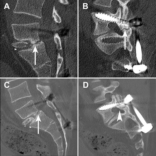
Septic nonunion after vertebral fusion can lead to significant patient disability. The management of septic nonunions usually involves surgical debridement, bone fixation, and antibiotic therapy. Particularly challenging is lumbosacral vertebral nonunions, which necessitate a difficult surgical approach. We present a novel approach using a scapula tip free flap through an intra-abdominal approach to reconstruct a L5–S1 vertebral defect after a septic nonunion. Our patient, 31-year-old man, with no medical conditions, had a fusion of L5–S1 due to severe lower back pain secondary to isthmic spondylolysis and spondylolisthesis. Despite multiple attempts of surgical fusion, postoperatively the patient developed a septic nonunion. Following a modified DAIR, the nonunion was reconstructed with a scapula tip bone flap 4 × 3 × 2 cm. The subscapular vessels were anastomosed to the deep inferior epigastric vessels after an intra-abdominal inset. The patient was discharged at 15 days postoperatively without any complications. At 1-year follow-up the patient is pain-free, off opiate analgesia with radiological evidence of fusion between the scapula tip, L5 and the S1 vertebral body. This case report describes the use, for the first time, of a free scapula tip, to a lumbosacral spinal defect. The use of the free scapula tip flap may be considered for reconstruction of osseous spinal defects due to its long pedicle and the unique bone shape.
期刊介绍:
Microsurgery is an international and interdisciplinary publication of original contributions concerning surgery under microscopic magnification. Microsurgery publishes clinical studies, research papers, invited articles, relevant reviews, and other scholarly works from all related fields including orthopaedic surgery, otolaryngology, pediatric surgery, plastic surgery, urology, and vascular surgery.


 分享
分享
 求助内容:
求助内容: 应助结果提醒方式:
 扫码关注我们
