{"title":"金属伪影减少工具和 mA 水平对使用锥束 CT 诊断牙髓治疗牙齿折断扩展的影响","authors":"Luísa Valente Gotardo Lara Alves, Cassiano Ricardo Ferreira Pires, Manoel Damião Sousa-Neto, Heitor Silva Prado, Jardel Francisco Mazzi-Chaves, Amanda Pelegrin Candemil","doi":"10.1007/s00784-024-05945-3","DOIUrl":null,"url":null,"abstract":"<h3 data-test=\"abstract-sub-heading\">Aim</h3><p>To evaluate the influence of different levels of metal artifact reduction (MAR) tool and milliamperage (mA) on the diagnosis of fracture extension in endodontically treated teeth using cone beam CT (CBCT).</p><h3 data-test=\"abstract-sub-heading\">Materials and methods</h3><p>Ten maxillary premolars were endodontically treated and positioned in the empty sockets of a human maxilla covered with wax. CBCT acquisitions were performed using the Eagle Edge device (Dabi Atlante, Brazil) adjusted to 120 kVp, FOV of 4 × 6 cm, exposure time of 24 s and voxel size of 0.2 mm in 8 different conditions with different MAR (1, 2 and 3) and mA (3.2 and 6.3) levels. Crown-root fractures were simulated in the universal testing machine, and CBCT images were acquired again. Five radiologists evaluated the presence and extension of fractures with a 5-point scale. Statistical analysis was performed by analysis of variance, Tukey and Kappa test (α = 0.05).</p><h3 data-test=\"abstract-sub-heading\">Results</h3><p>Although different mA levels did not significantly (<i>p</i> > 0.05) affect the diagnosis values for fracture presence and extension, when evaluated the different levels of MAR, AUC and sensitivity showed significantly higher values (<i>p</i> < 0.05) for MAR 0 using 6.3 mA and kappa agreement showed significantly higher values (<i>p</i> < 0.05) for MAR 0 and 2 using 6.3 mA.</p><h3 data-test=\"abstract-sub-heading\">Conclusions</h3><p>Although mA levels do not have a diagnostic effect when isolating the MAR level; in 6.3 mA, MAR 0 and 2 can positively influence the diagnosis of fracture extension in endodontically treated teeth using CBCT.</p><h3 data-test=\"abstract-sub-heading\">Clinical relevance</h3><p>The isolate evaluation of dental fracture presence can overlook diagnostics error of its extension.</p>","PeriodicalId":10461,"journal":{"name":"Clinical Oral Investigations","volume":"28 1","pages":""},"PeriodicalIF":3.6000,"publicationDate":"2024-09-19","publicationTypes":"Journal Article","fieldsOfStudy":null,"isOpenAccess":false,"openAccessPdf":"","citationCount":"0","resultStr":"{\"title\":\"Metal artifact reduction tool and mA levels impact on the diagnosis of fracture extension in endodontically treated teeth using cone-beam CT\",\"authors\":\"Luísa Valente Gotardo Lara Alves, Cassiano Ricardo Ferreira Pires, Manoel Damião Sousa-Neto, Heitor Silva Prado, Jardel Francisco Mazzi-Chaves, Amanda Pelegrin Candemil\",\"doi\":\"10.1007/s00784-024-05945-3\",\"DOIUrl\":null,\"url\":null,\"abstract\":\"<h3 data-test=\\\"abstract-sub-heading\\\">Aim</h3><p>To evaluate the influence of different levels of metal artifact reduction (MAR) tool and milliamperage (mA) on the diagnosis of fracture extension in endodontically treated teeth using cone beam CT (CBCT).</p><h3 data-test=\\\"abstract-sub-heading\\\">Materials and methods</h3><p>Ten maxillary premolars were endodontically treated and positioned in the empty sockets of a human maxilla covered with wax. CBCT acquisitions were performed using the Eagle Edge device (Dabi Atlante, Brazil) adjusted to 120 kVp, FOV of 4 × 6 cm, exposure time of 24 s and voxel size of 0.2 mm in 8 different conditions with different MAR (1, 2 and 3) and mA (3.2 and 6.3) levels. Crown-root fractures were simulated in the universal testing machine, and CBCT images were acquired again. Five radiologists evaluated the presence and extension of fractures with a 5-point scale. Statistical analysis was performed by analysis of variance, Tukey and Kappa test (α = 0.05).</p><h3 data-test=\\\"abstract-sub-heading\\\">Results</h3><p>Although different mA levels did not significantly (<i>p</i> > 0.05) affect the diagnosis values for fracture presence and extension, when evaluated the different levels of MAR, AUC and sensitivity showed significantly higher values (<i>p</i> < 0.05) for MAR 0 using 6.3 mA and kappa agreement showed significantly higher values (<i>p</i> < 0.05) for MAR 0 and 2 using 6.3 mA.</p><h3 data-test=\\\"abstract-sub-heading\\\">Conclusions</h3><p>Although mA levels do not have a diagnostic effect when isolating the MAR level; in 6.3 mA, MAR 0 and 2 can positively influence the diagnosis of fracture extension in endodontically treated teeth using CBCT.</p><h3 data-test=\\\"abstract-sub-heading\\\">Clinical relevance</h3><p>The isolate evaluation of dental fracture presence can overlook diagnostics error of its extension.</p>\",\"PeriodicalId\":10461,\"journal\":{\"name\":\"Clinical Oral Investigations\",\"volume\":\"28 1\",\"pages\":\"\"},\"PeriodicalIF\":3.6000,\"publicationDate\":\"2024-09-19\",\"publicationTypes\":\"Journal Article\",\"fieldsOfStudy\":null,\"isOpenAccess\":false,\"openAccessPdf\":\"\",\"citationCount\":\"0\",\"resultStr\":null,\"platform\":\"Semanticscholar\",\"paperid\":null,\"PeriodicalName\":\"Clinical Oral Investigations\",\"FirstCategoryId\":\"3\",\"ListUrlMain\":\"https://doi.org/10.1007/s00784-024-05945-3\",\"RegionNum\":2,\"RegionCategory\":\"医学\",\"ArticlePicture\":[],\"TitleCN\":null,\"AbstractTextCN\":null,\"PMCID\":null,\"EPubDate\":\"\",\"PubModel\":\"\",\"JCR\":\"Q1\",\"JCRName\":\"DENTISTRY, ORAL SURGERY & MEDICINE\",\"Score\":null,\"Total\":0}","platform":"Semanticscholar","paperid":null,"PeriodicalName":"Clinical Oral Investigations","FirstCategoryId":"3","ListUrlMain":"https://doi.org/10.1007/s00784-024-05945-3","RegionNum":2,"RegionCategory":"医学","ArticlePicture":[],"TitleCN":null,"AbstractTextCN":null,"PMCID":null,"EPubDate":"","PubModel":"","JCR":"Q1","JCRName":"DENTISTRY, ORAL SURGERY & MEDICINE","Score":null,"Total":0}
Metal artifact reduction tool and mA levels impact on the diagnosis of fracture extension in endodontically treated teeth using cone-beam CT
Aim
To evaluate the influence of different levels of metal artifact reduction (MAR) tool and milliamperage (mA) on the diagnosis of fracture extension in endodontically treated teeth using cone beam CT (CBCT).
Materials and methods
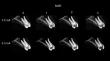
Ten maxillary premolars were endodontically treated and positioned in the empty sockets of a human maxilla covered with wax. CBCT acquisitions were performed using the Eagle Edge device (Dabi Atlante, Brazil) adjusted to 120 kVp, FOV of 4 × 6 cm, exposure time of 24 s and voxel size of 0.2 mm in 8 different conditions with different MAR (1, 2 and 3) and mA (3.2 and 6.3) levels. Crown-root fractures were simulated in the universal testing machine, and CBCT images were acquired again. Five radiologists evaluated the presence and extension of fractures with a 5-point scale. Statistical analysis was performed by analysis of variance, Tukey and Kappa test (α = 0.05).
Results
Although different mA levels did not significantly (p > 0.05) affect the diagnosis values for fracture presence and extension, when evaluated the different levels of MAR, AUC and sensitivity showed significantly higher values (p < 0.05) for MAR 0 using 6.3 mA and kappa agreement showed significantly higher values (p < 0.05) for MAR 0 and 2 using 6.3 mA.
Conclusions
Although mA levels do not have a diagnostic effect when isolating the MAR level; in 6.3 mA, MAR 0 and 2 can positively influence the diagnosis of fracture extension in endodontically treated teeth using CBCT.
Clinical relevance
The isolate evaluation of dental fracture presence can overlook diagnostics error of its extension.
期刊介绍:
The journal Clinical Oral Investigations is a multidisciplinary, international forum for publication of research from all fields of oral medicine. The journal publishes original scientific articles and invited reviews which provide up-to-date results of basic and clinical studies in oral and maxillofacial science and medicine. The aim is to clarify the relevance of new results to modern practice, for an international readership. Coverage includes maxillofacial and oral surgery, prosthetics and restorative dentistry, operative dentistry, endodontics, periodontology, orthodontics, dental materials science, clinical trials, epidemiology, pedodontics, oral implant, preventive dentistiry, oral pathology, oral basic sciences and more.


 分享
分享
 求助内容:
求助内容: 应助结果提醒方式:
 扫码关注我们
