Jatin Singh, Grant Kokenberger, Lucas Pu, Ernest Chan, Alaa Ali, Kaveh Moghbeli, Tong Yu, Chadi A. Hage, Pablo G. Sanchez, Jiantao Pu
{"title":"利用术前胸部 CT 扫描的 CT 衍生特征预测系统性硬化症患者肺移植后的存活率","authors":"Jatin Singh, Grant Kokenberger, Lucas Pu, Ernest Chan, Alaa Ali, Kaveh Moghbeli, Tong Yu, Chadi A. Hage, Pablo G. Sanchez, Jiantao Pu","doi":"10.1007/s00330-024-11077-9","DOIUrl":null,"url":null,"abstract":"<h3 data-test=\"abstract-sub-heading\">Objectives</h3><p>The current understanding of survival prediction of lung transplant (LTx) patients with systemic sclerosis (SSc) is limited. This study aims to identify novel image features from preoperative chest CT scans associated with post-LTx survival in SSc patients and integrate them into comprehensive prediction models.</p><h3 data-test=\"abstract-sub-heading\">Materials and methods</h3><p>We conducted a retrospective study based on a cohort of SSc patients with demographic information, clinical data, and preoperative chest CT scans who underwent LTx between 2004 and 2020. This cohort consists of 102 patients (mean age, 50 years ± 10, 61% (62/102) females). Five CT-derived body composition features (bone, skeletal muscle, visceral, subcutaneous, and intramuscular adipose tissues) and three CT-derived cardiopulmonary features (heart, arteries, and veins) were automatically computed using 3-D convolutional neural networks. Cox regression was used to identify post-LTx survival factors, generate composite prediction models, and stratify patients based on mortality risk. Model performance was assessed using the area under the receiver operating characteristics curve (ROC-AUC).</p><h3 data-test=\"abstract-sub-heading\">Results</h3><p>Muscle mass ratio, bone density, artery–vein volume ratio, muscle volume, and heart volume ratio computed from CT images were significantly associated with post-LTx survival. Models using only CT-derived features outperformed all state-of-the-art clinical models in predicting post-LTx survival. The addition of CT-derived features improved the performance of traditional models at 1-year, 3-year, and 5-year survival prediction with maximum AUC scores of 0.77 (0.67–0.86), 0.85 (0.77–0.93), and 0.90 (95% CI: 0.83–0.97), respectively.</p><h3 data-test=\"abstract-sub-heading\">Conclusion</h3><p>The integration of CT-derived features with demographic and clinical features can significantly improve t post-LTx survival prediction and identify high-risk SSc patients.</p><h3 data-test=\"abstract-sub-heading\">Key Points</h3><p><b><i>Question</i></b> <i>What CT features can predict post-lung-transplant survival for SSc patients</i>?</p><p><b><i>Finding</i></b> <i>CT body composition features such as muscle mass, bone density, and cardiopulmonary volumes significantly predict survival</i>.</p><p><b><i>Clinical relevance</i></b> <i>Our individualized risk assessment tool can better guide clinicians in choosing and managing patients requiring lung transplant for systemic sclerosis</i>.</p><h3 data-test=\"abstract-sub-heading\">Graphical Abstract</h3>","PeriodicalId":12076,"journal":{"name":"European Radiology","volume":"20 1","pages":""},"PeriodicalIF":4.7000,"publicationDate":"2024-09-18","publicationTypes":"Journal Article","fieldsOfStudy":null,"isOpenAccess":false,"openAccessPdf":"","citationCount":"0","resultStr":"{\"title\":\"Predicting post-lung transplant survival in systemic sclerosis using CT-derived features from preoperative chest CT scans\",\"authors\":\"Jatin Singh, Grant Kokenberger, Lucas Pu, Ernest Chan, Alaa Ali, Kaveh Moghbeli, Tong Yu, Chadi A. Hage, Pablo G. Sanchez, Jiantao Pu\",\"doi\":\"10.1007/s00330-024-11077-9\",\"DOIUrl\":null,\"url\":null,\"abstract\":\"<h3 data-test=\\\"abstract-sub-heading\\\">Objectives</h3><p>The current understanding of survival prediction of lung transplant (LTx) patients with systemic sclerosis (SSc) is limited. This study aims to identify novel image features from preoperative chest CT scans associated with post-LTx survival in SSc patients and integrate them into comprehensive prediction models.</p><h3 data-test=\\\"abstract-sub-heading\\\">Materials and methods</h3><p>We conducted a retrospective study based on a cohort of SSc patients with demographic information, clinical data, and preoperative chest CT scans who underwent LTx between 2004 and 2020. This cohort consists of 102 patients (mean age, 50 years ± 10, 61% (62/102) females). Five CT-derived body composition features (bone, skeletal muscle, visceral, subcutaneous, and intramuscular adipose tissues) and three CT-derived cardiopulmonary features (heart, arteries, and veins) were automatically computed using 3-D convolutional neural networks. Cox regression was used to identify post-LTx survival factors, generate composite prediction models, and stratify patients based on mortality risk. Model performance was assessed using the area under the receiver operating characteristics curve (ROC-AUC).</p><h3 data-test=\\\"abstract-sub-heading\\\">Results</h3><p>Muscle mass ratio, bone density, artery–vein volume ratio, muscle volume, and heart volume ratio computed from CT images were significantly associated with post-LTx survival. Models using only CT-derived features outperformed all state-of-the-art clinical models in predicting post-LTx survival. The addition of CT-derived features improved the performance of traditional models at 1-year, 3-year, and 5-year survival prediction with maximum AUC scores of 0.77 (0.67–0.86), 0.85 (0.77–0.93), and 0.90 (95% CI: 0.83–0.97), respectively.</p><h3 data-test=\\\"abstract-sub-heading\\\">Conclusion</h3><p>The integration of CT-derived features with demographic and clinical features can significantly improve t post-LTx survival prediction and identify high-risk SSc patients.</p><h3 data-test=\\\"abstract-sub-heading\\\">Key Points</h3><p><b><i>Question</i></b> <i>What CT features can predict post-lung-transplant survival for SSc patients</i>?</p><p><b><i>Finding</i></b> <i>CT body composition features such as muscle mass, bone density, and cardiopulmonary volumes significantly predict survival</i>.</p><p><b><i>Clinical relevance</i></b> <i>Our individualized risk assessment tool can better guide clinicians in choosing and managing patients requiring lung transplant for systemic sclerosis</i>.</p><h3 data-test=\\\"abstract-sub-heading\\\">Graphical Abstract</h3>\",\"PeriodicalId\":12076,\"journal\":{\"name\":\"European Radiology\",\"volume\":\"20 1\",\"pages\":\"\"},\"PeriodicalIF\":4.7000,\"publicationDate\":\"2024-09-18\",\"publicationTypes\":\"Journal Article\",\"fieldsOfStudy\":null,\"isOpenAccess\":false,\"openAccessPdf\":\"\",\"citationCount\":\"0\",\"resultStr\":null,\"platform\":\"Semanticscholar\",\"paperid\":null,\"PeriodicalName\":\"European Radiology\",\"FirstCategoryId\":\"3\",\"ListUrlMain\":\"https://doi.org/10.1007/s00330-024-11077-9\",\"RegionNum\":2,\"RegionCategory\":\"医学\",\"ArticlePicture\":[],\"TitleCN\":null,\"AbstractTextCN\":null,\"PMCID\":null,\"EPubDate\":\"\",\"PubModel\":\"\",\"JCR\":\"Q1\",\"JCRName\":\"RADIOLOGY, NUCLEAR MEDICINE & MEDICAL IMAGING\",\"Score\":null,\"Total\":0}","platform":"Semanticscholar","paperid":null,"PeriodicalName":"European Radiology","FirstCategoryId":"3","ListUrlMain":"https://doi.org/10.1007/s00330-024-11077-9","RegionNum":2,"RegionCategory":"医学","ArticlePicture":[],"TitleCN":null,"AbstractTextCN":null,"PMCID":null,"EPubDate":"","PubModel":"","JCR":"Q1","JCRName":"RADIOLOGY, NUCLEAR MEDICINE & MEDICAL IMAGING","Score":null,"Total":0}
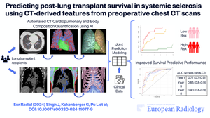
Predicting post-lung transplant survival in systemic sclerosis using CT-derived features from preoperative chest CT scans
Objectives
The current understanding of survival prediction of lung transplant (LTx) patients with systemic sclerosis (SSc) is limited. This study aims to identify novel image features from preoperative chest CT scans associated with post-LTx survival in SSc patients and integrate them into comprehensive prediction models.
Materials and methods
We conducted a retrospective study based on a cohort of SSc patients with demographic information, clinical data, and preoperative chest CT scans who underwent LTx between 2004 and 2020. This cohort consists of 102 patients (mean age, 50 years ± 10, 61% (62/102) females). Five CT-derived body composition features (bone, skeletal muscle, visceral, subcutaneous, and intramuscular adipose tissues) and three CT-derived cardiopulmonary features (heart, arteries, and veins) were automatically computed using 3-D convolutional neural networks. Cox regression was used to identify post-LTx survival factors, generate composite prediction models, and stratify patients based on mortality risk. Model performance was assessed using the area under the receiver operating characteristics curve (ROC-AUC).
Results
Muscle mass ratio, bone density, artery–vein volume ratio, muscle volume, and heart volume ratio computed from CT images were significantly associated with post-LTx survival. Models using only CT-derived features outperformed all state-of-the-art clinical models in predicting post-LTx survival. The addition of CT-derived features improved the performance of traditional models at 1-year, 3-year, and 5-year survival prediction with maximum AUC scores of 0.77 (0.67–0.86), 0.85 (0.77–0.93), and 0.90 (95% CI: 0.83–0.97), respectively.
Conclusion
The integration of CT-derived features with demographic and clinical features can significantly improve t post-LTx survival prediction and identify high-risk SSc patients.
Key Points
QuestionWhat CT features can predict post-lung-transplant survival for SSc patients?
FindingCT body composition features such as muscle mass, bone density, and cardiopulmonary volumes significantly predict survival.
Clinical relevanceOur individualized risk assessment tool can better guide clinicians in choosing and managing patients requiring lung transplant for systemic sclerosis.
期刊介绍:
European Radiology (ER) continuously updates scientific knowledge in radiology by publication of strong original articles and state-of-the-art reviews written by leading radiologists. A well balanced combination of review articles, original papers, short communications from European radiological congresses and information on society matters makes ER an indispensable source for current information in this field.
This is the Journal of the European Society of Radiology, and the official journal of a number of societies.
From 2004-2008 supplements to European Radiology were published under its companion, European Radiology Supplements, ISSN 1613-3749.


 分享
分享
 求助内容:
求助内容: 应助结果提醒方式:
 扫码关注我们
