Gabrielle C. Colleran, Maria Fossmark, Karen Rosendahl, Maria Argyropoulou, Kshitij Mankad, Amaka C. Offiah
{"title":"ESR要点:疑似虐待儿童的成像--欧洲儿科放射学会的实践建议","authors":"Gabrielle C. Colleran, Maria Fossmark, Karen Rosendahl, Maria Argyropoulou, Kshitij Mankad, Amaka C. Offiah","doi":"10.1007/s00330-024-11052-4","DOIUrl":null,"url":null,"abstract":"<h3 data-test=\"abstract-sub-heading\">Abstract</h3><p>The goal of this paper is to provide a useful desktop reference for the imaging of suspected child abuse with clear, age-specific pathways for appropriate evidence-based imaging and follow-up.</p><p>We aim to provide a road map for the imaging evaluation and follow-up of this important and vulnerable cohort of patients presenting with signs and symptoms concerning for inflicted injury. As the imaging recommendations differ for children of different ages, we provide a flowchart of the appropriate imaging pathway for infants, toddlers, and older children, which allows ease of selection of which children should undergo skeletal survey, non-contrast computed tomography (CT) brain with 3-dimensional (D) reformats, and magnetic resonance imaging (MRI) of the brain and whole spine. For ease of review, we include a table of the common intracranial and spinal patterns of injury in abusive head trauma. We summarise search patterns, areas of review, and key findings to include in the report.</p><p>To exclude skeletal injury, infants and children under 2 years of age should undergo a full skeletal survey in accordance with national guidelines, with a limited follow-up skeletal survey performed 11–14 days later. For children over 2 years of age, the need for skeletal imaging should be decided on a case-by-case basis.</p><p>All infants should undergo a non-contrast-enhanced CT brain with 3-D reformats. If this is normal with no abnormal neurology, then no further neuroimaging is required. If this is abnormal, then they should proceed to MRI brain and whole spine within 2–5 days. Children older than 1 year of age who have abnormal neurology and/or findings on skeletal survey that are suggestive of inflicted injury should undergo non-contrast CT brain with 3-D reformats and, depending on the findings, may also require MRI of the brain and whole spine.</p><p>We hope that this will be a helpful contribution to the radiology literature, particularly for the general radiologist with low volumes of paediatrics in their practice, supporting them with managing these important cases when they arise in daily practice.</p><h3 data-test=\"abstract-sub-heading\">Key Points</h3><ul>\n<li>\n<p><i>The choice of initial imaging (skeletal survey and/or brain CT) depends on the age of the child in whom abuse is suspected</i>.</p>\n</li>\n<li>\n<p><i>A follow-up skeletal survey is mandatory 11–14 days after the initial survey</i>.</p>\n</li>\n<li>\n<p><i>If an MRI of the brain is performed, then an MRI of the whole spine should be performed concurrently</i>.</p>\n</li>\n</ul>","PeriodicalId":12076,"journal":{"name":"European Radiology","volume":"48 1","pages":""},"PeriodicalIF":4.7000,"publicationDate":"2024-09-18","publicationTypes":"Journal Article","fieldsOfStudy":null,"isOpenAccess":false,"openAccessPdf":"","citationCount":"0","resultStr":"{\"title\":\"ESR Essentials: imaging of suspected child abuse—practice recommendations by the European Society of Paediatric Radiology\",\"authors\":\"Gabrielle C. Colleran, Maria Fossmark, Karen Rosendahl, Maria Argyropoulou, Kshitij Mankad, Amaka C. Offiah\",\"doi\":\"10.1007/s00330-024-11052-4\",\"DOIUrl\":null,\"url\":null,\"abstract\":\"<h3 data-test=\\\"abstract-sub-heading\\\">Abstract</h3><p>The goal of this paper is to provide a useful desktop reference for the imaging of suspected child abuse with clear, age-specific pathways for appropriate evidence-based imaging and follow-up.</p><p>We aim to provide a road map for the imaging evaluation and follow-up of this important and vulnerable cohort of patients presenting with signs and symptoms concerning for inflicted injury. As the imaging recommendations differ for children of different ages, we provide a flowchart of the appropriate imaging pathway for infants, toddlers, and older children, which allows ease of selection of which children should undergo skeletal survey, non-contrast computed tomography (CT) brain with 3-dimensional (D) reformats, and magnetic resonance imaging (MRI) of the brain and whole spine. For ease of review, we include a table of the common intracranial and spinal patterns of injury in abusive head trauma. We summarise search patterns, areas of review, and key findings to include in the report.</p><p>To exclude skeletal injury, infants and children under 2 years of age should undergo a full skeletal survey in accordance with national guidelines, with a limited follow-up skeletal survey performed 11–14 days later. For children over 2 years of age, the need for skeletal imaging should be decided on a case-by-case basis.</p><p>All infants should undergo a non-contrast-enhanced CT brain with 3-D reformats. If this is normal with no abnormal neurology, then no further neuroimaging is required. If this is abnormal, then they should proceed to MRI brain and whole spine within 2–5 days. Children older than 1 year of age who have abnormal neurology and/or findings on skeletal survey that are suggestive of inflicted injury should undergo non-contrast CT brain with 3-D reformats and, depending on the findings, may also require MRI of the brain and whole spine.</p><p>We hope that this will be a helpful contribution to the radiology literature, particularly for the general radiologist with low volumes of paediatrics in their practice, supporting them with managing these important cases when they arise in daily practice.</p><h3 data-test=\\\"abstract-sub-heading\\\">Key Points</h3><ul>\\n<li>\\n<p><i>The choice of initial imaging (skeletal survey and/or brain CT) depends on the age of the child in whom abuse is suspected</i>.</p>\\n</li>\\n<li>\\n<p><i>A follow-up skeletal survey is mandatory 11–14 days after the initial survey</i>.</p>\\n</li>\\n<li>\\n<p><i>If an MRI of the brain is performed, then an MRI of the whole spine should be performed concurrently</i>.</p>\\n</li>\\n</ul>\",\"PeriodicalId\":12076,\"journal\":{\"name\":\"European Radiology\",\"volume\":\"48 1\",\"pages\":\"\"},\"PeriodicalIF\":4.7000,\"publicationDate\":\"2024-09-18\",\"publicationTypes\":\"Journal Article\",\"fieldsOfStudy\":null,\"isOpenAccess\":false,\"openAccessPdf\":\"\",\"citationCount\":\"0\",\"resultStr\":null,\"platform\":\"Semanticscholar\",\"paperid\":null,\"PeriodicalName\":\"European Radiology\",\"FirstCategoryId\":\"3\",\"ListUrlMain\":\"https://doi.org/10.1007/s00330-024-11052-4\",\"RegionNum\":2,\"RegionCategory\":\"医学\",\"ArticlePicture\":[],\"TitleCN\":null,\"AbstractTextCN\":null,\"PMCID\":null,\"EPubDate\":\"\",\"PubModel\":\"\",\"JCR\":\"Q1\",\"JCRName\":\"RADIOLOGY, NUCLEAR MEDICINE & MEDICAL IMAGING\",\"Score\":null,\"Total\":0}","platform":"Semanticscholar","paperid":null,"PeriodicalName":"European Radiology","FirstCategoryId":"3","ListUrlMain":"https://doi.org/10.1007/s00330-024-11052-4","RegionNum":2,"RegionCategory":"医学","ArticlePicture":[],"TitleCN":null,"AbstractTextCN":null,"PMCID":null,"EPubDate":"","PubModel":"","JCR":"Q1","JCRName":"RADIOLOGY, NUCLEAR MEDICINE & MEDICAL IMAGING","Score":null,"Total":0}
ESR Essentials: imaging of suspected child abuse—practice recommendations by the European Society of Paediatric Radiology
Abstract
The goal of this paper is to provide a useful desktop reference for the imaging of suspected child abuse with clear, age-specific pathways for appropriate evidence-based imaging and follow-up.
We aim to provide a road map for the imaging evaluation and follow-up of this important and vulnerable cohort of patients presenting with signs and symptoms concerning for inflicted injury. As the imaging recommendations differ for children of different ages, we provide a flowchart of the appropriate imaging pathway for infants, toddlers, and older children, which allows ease of selection of which children should undergo skeletal survey, non-contrast computed tomography (CT) brain with 3-dimensional (D) reformats, and magnetic resonance imaging (MRI) of the brain and whole spine. For ease of review, we include a table of the common intracranial and spinal patterns of injury in abusive head trauma. We summarise search patterns, areas of review, and key findings to include in the report.
To exclude skeletal injury, infants and children under 2 years of age should undergo a full skeletal survey in accordance with national guidelines, with a limited follow-up skeletal survey performed 11–14 days later. For children over 2 years of age, the need for skeletal imaging should be decided on a case-by-case basis.
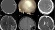
All infants should undergo a non-contrast-enhanced CT brain with 3-D reformats. If this is normal with no abnormal neurology, then no further neuroimaging is required. If this is abnormal, then they should proceed to MRI brain and whole spine within 2–5 days. Children older than 1 year of age who have abnormal neurology and/or findings on skeletal survey that are suggestive of inflicted injury should undergo non-contrast CT brain with 3-D reformats and, depending on the findings, may also require MRI of the brain and whole spine.
We hope that this will be a helpful contribution to the radiology literature, particularly for the general radiologist with low volumes of paediatrics in their practice, supporting them with managing these important cases when they arise in daily practice.
Key Points
The choice of initial imaging (skeletal survey and/or brain CT) depends on the age of the child in whom abuse is suspected.
A follow-up skeletal survey is mandatory 11–14 days after the initial survey.
If an MRI of the brain is performed, then an MRI of the whole spine should be performed concurrently.
期刊介绍:
European Radiology (ER) continuously updates scientific knowledge in radiology by publication of strong original articles and state-of-the-art reviews written by leading radiologists. A well balanced combination of review articles, original papers, short communications from European radiological congresses and information on society matters makes ER an indispensable source for current information in this field.
This is the Journal of the European Society of Radiology, and the official journal of a number of societies.
From 2004-2008 supplements to European Radiology were published under its companion, European Radiology Supplements, ISSN 1613-3749.


 分享
分享
 求助内容:
求助内容: 应助结果提醒方式:
 扫码关注我们
