John A Ashindoitiang, Victor I Canice Nwagbara, Ekpo E Edet, Theophilus Ipeh Ugbem, Joseph S Ukam, Maurice E Asuquo
{"title":"表现为腹腔内肿瘤的巨大浆膜下子宫肌瘤:病例报告。","authors":"John A Ashindoitiang, Victor I Canice Nwagbara, Ekpo E Edet, Theophilus Ipeh Ugbem, Joseph S Ukam, Maurice E Asuquo","doi":"10.1177/20363613241285089","DOIUrl":null,"url":null,"abstract":"<p><p>Uterine leiomyomas are common benign gynecological tumors due to the overgrowth of uterine smooth muscle. Pedunculated uterine leiomyoma occurs when the mass is in continuity with the uterus with a stalk and may grow either within the uterine cavity or outside of the uterus and may mimic ovarian neoplasms or intraabdominal tumors. Presented is a 28-year-old woman with a progressive abdominal swelling in the past 9 months seen at the surgical outpatient of our facility. Preoperative CT suggested a diagnosis of an intrabdominal cystic. She had laparotomy and was offered myomectomies on account of a large subserous uterine mass arising from the right side of the uterine fundus, small subserous fundal mass, intramural mass in the left side of the fundus and a cervical mass. Histology confirmed multiple uterine leiomyomas with extensive cystic degenerative changes of the large subserous uterine myoma and adenomyosis of the left fundal mass. Detecting the continuity of an abdominal mass even with extensive degenerative changes mimicking a cyst in continuity with the uterus by a pedicle sign on imaging in the absence of ascites should arouse the diagnosis of pedunculated subserosal leiomyoma. This should be further heightened when it is found in association with cervical myoma. Subserous uterine leiomyoma should be considered in a patient of childbearing age with a grossly distended abdomen without obvious evidence of pregnancy or malignancy. Large subserous uterine leiomyoma in an intraabdominal location may present with diagnostic and surgical challenges that require interdisciplinary cooperation.</p>","PeriodicalId":46078,"journal":{"name":"Rare Tumors","volume":"16 ","pages":"20363613241285089"},"PeriodicalIF":0.9000,"publicationDate":"2024-09-12","publicationTypes":"Journal Article","fieldsOfStudy":null,"isOpenAccess":false,"openAccessPdf":"https://www.ncbi.nlm.nih.gov/pmc/articles/PMC11406654/pdf/","citationCount":"0","resultStr":"{\"title\":\"Large subserous uterine leiomyoma presenting as intraabdominal tumor: A case report.\",\"authors\":\"John A Ashindoitiang, Victor I Canice Nwagbara, Ekpo E Edet, Theophilus Ipeh Ugbem, Joseph S Ukam, Maurice E Asuquo\",\"doi\":\"10.1177/20363613241285089\",\"DOIUrl\":null,\"url\":null,\"abstract\":\"<p><p>Uterine leiomyomas are common benign gynecological tumors due to the overgrowth of uterine smooth muscle. Pedunculated uterine leiomyoma occurs when the mass is in continuity with the uterus with a stalk and may grow either within the uterine cavity or outside of the uterus and may mimic ovarian neoplasms or intraabdominal tumors. Presented is a 28-year-old woman with a progressive abdominal swelling in the past 9 months seen at the surgical outpatient of our facility. Preoperative CT suggested a diagnosis of an intrabdominal cystic. She had laparotomy and was offered myomectomies on account of a large subserous uterine mass arising from the right side of the uterine fundus, small subserous fundal mass, intramural mass in the left side of the fundus and a cervical mass. Histology confirmed multiple uterine leiomyomas with extensive cystic degenerative changes of the large subserous uterine myoma and adenomyosis of the left fundal mass. Detecting the continuity of an abdominal mass even with extensive degenerative changes mimicking a cyst in continuity with the uterus by a pedicle sign on imaging in the absence of ascites should arouse the diagnosis of pedunculated subserosal leiomyoma. This should be further heightened when it is found in association with cervical myoma. Subserous uterine leiomyoma should be considered in a patient of childbearing age with a grossly distended abdomen without obvious evidence of pregnancy or malignancy. Large subserous uterine leiomyoma in an intraabdominal location may present with diagnostic and surgical challenges that require interdisciplinary cooperation.</p>\",\"PeriodicalId\":46078,\"journal\":{\"name\":\"Rare Tumors\",\"volume\":\"16 \",\"pages\":\"20363613241285089\"},\"PeriodicalIF\":0.9000,\"publicationDate\":\"2024-09-12\",\"publicationTypes\":\"Journal Article\",\"fieldsOfStudy\":null,\"isOpenAccess\":false,\"openAccessPdf\":\"https://www.ncbi.nlm.nih.gov/pmc/articles/PMC11406654/pdf/\",\"citationCount\":\"0\",\"resultStr\":null,\"platform\":\"Semanticscholar\",\"paperid\":null,\"PeriodicalName\":\"Rare Tumors\",\"FirstCategoryId\":\"1085\",\"ListUrlMain\":\"https://doi.org/10.1177/20363613241285089\",\"RegionNum\":0,\"RegionCategory\":null,\"ArticlePicture\":[],\"TitleCN\":null,\"AbstractTextCN\":null,\"PMCID\":null,\"EPubDate\":\"2024/1/1 0:00:00\",\"PubModel\":\"eCollection\",\"JCR\":\"Q4\",\"JCRName\":\"ONCOLOGY\",\"Score\":null,\"Total\":0}","platform":"Semanticscholar","paperid":null,"PeriodicalName":"Rare Tumors","FirstCategoryId":"1085","ListUrlMain":"https://doi.org/10.1177/20363613241285089","RegionNum":0,"RegionCategory":null,"ArticlePicture":[],"TitleCN":null,"AbstractTextCN":null,"PMCID":null,"EPubDate":"2024/1/1 0:00:00","PubModel":"eCollection","JCR":"Q4","JCRName":"ONCOLOGY","Score":null,"Total":0}
Large subserous uterine leiomyoma presenting as intraabdominal tumor: A case report.
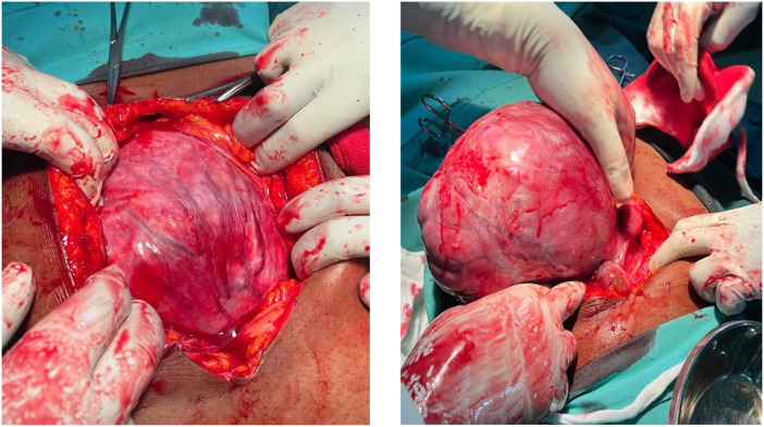
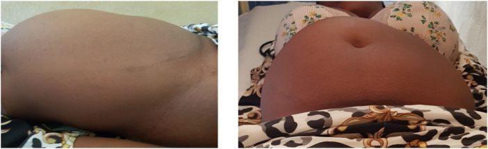
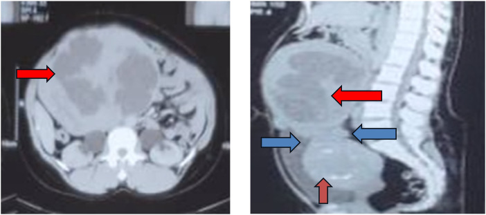
Uterine leiomyomas are common benign gynecological tumors due to the overgrowth of uterine smooth muscle. Pedunculated uterine leiomyoma occurs when the mass is in continuity with the uterus with a stalk and may grow either within the uterine cavity or outside of the uterus and may mimic ovarian neoplasms or intraabdominal tumors. Presented is a 28-year-old woman with a progressive abdominal swelling in the past 9 months seen at the surgical outpatient of our facility. Preoperative CT suggested a diagnosis of an intrabdominal cystic. She had laparotomy and was offered myomectomies on account of a large subserous uterine mass arising from the right side of the uterine fundus, small subserous fundal mass, intramural mass in the left side of the fundus and a cervical mass. Histology confirmed multiple uterine leiomyomas with extensive cystic degenerative changes of the large subserous uterine myoma and adenomyosis of the left fundal mass. Detecting the continuity of an abdominal mass even with extensive degenerative changes mimicking a cyst in continuity with the uterus by a pedicle sign on imaging in the absence of ascites should arouse the diagnosis of pedunculated subserosal leiomyoma. This should be further heightened when it is found in association with cervical myoma. Subserous uterine leiomyoma should be considered in a patient of childbearing age with a grossly distended abdomen without obvious evidence of pregnancy or malignancy. Large subserous uterine leiomyoma in an intraabdominal location may present with diagnostic and surgical challenges that require interdisciplinary cooperation.


 分享
分享
 求助内容:
求助内容: 应助结果提醒方式:
 扫码关注我们
