下载PDF
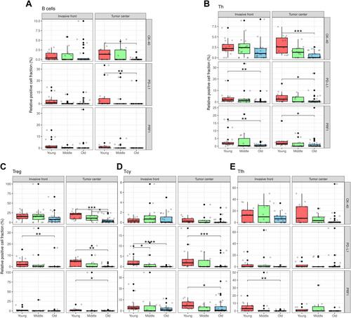
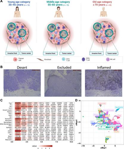
{"title":"剖析原发性管腔 B 型乳腺癌的免疫浸润与年龄的关系。","authors":"Sigrid Hatse, Yentl Lambrechts, Asier Antoranz Martinez, Maxim De Schepper, Tatjana Geukens, Hanne Vos, Lieze Berben, Julie Messiaen, Lukas Marcelis, Yannick Van Herck, Patrick Neven, Ann Smeets, Christine Desmedt, Frederik De Smet, Francesca Maria Bosisio, Hans Wildiers, Giuseppe Floris","doi":"10.1002/path.6354","DOIUrl":null,"url":null,"abstract":"<p>The impact of aging on the immune landscape of luminal breast cancer (Lum-BC) is poorly characterized. Understanding the age-related dynamics of immune editing in Lum-BC is anticipated to improve the therapeutic benefit of immunotherapy in older patients. To this end, here we applied the ‘multiple iterative labeling by antibody neo-deposition’ (MILAN) technique, a spatially resolved single-cell multiplex immunohistochemistry method. We created tissue microarrays by sampling both the tumor center and invasive front of luminal breast tumors collected from a cohort of treatment-naïve patients enrolled in the prospective monocentric IMAGE (IMmune system and AGEing) study. Patients were subdivided into three nonoverlapping age categories (35–45 = ‘young’, <i>n</i> = 12; 55–65 = ‘middle’, <i>n</i> = 15; ≥70 = ‘old’, <i>n</i> = 26). Additionally, depending on localization and amount of cytotoxic T lymphocytes, the tumor immune types ‘desert’ (<i>n</i> = 22), ‘excluded’ (<i>n</i> = 19), and ‘inflamed’ (<i>n</i> = 12) were identified. For the MILAN technique we used 58 markers comprising phenotypic and functional markers allowing in-depth characterization of T and B lymphocytes (T&B-lym). These were compared between age groups and tumor immune types using Wilcoxon's test and Pearson's correlation. Cytometric analysis revealed a decline of the immune cell compartment with aging. T&B-lym were numerically less abundant in tumors from middle-aged and old compared to young patients, regardless of the geographical tumor zone. Likewise, desert-type tumors showed the smallest immune-cell compartment and were not represented in the group of young patients. Analysis of immune checkpoint molecules revealed a heterogeneous geographical pattern of expression, indicating higher numbers of PD-L1 and OX40-positive T&B-lym in young compared to old patients. Despite the numerical decline of immune infiltration, old patients retained higher expression levels of OX40 in T helper cells located near cancer cells, compared to middle-aged and young patients. Aging is associated with important numerical and functional changes of the immune landscape in Lum-BC. © 2024 The Author(s). <i>The Journal of Pathology</i> published by John Wiley & Sons Ltd on behalf of The Pathological Society of Great Britain and Ireland.</p>","PeriodicalId":232,"journal":{"name":"The Journal of Pathology","volume":"264 3","pages":"344-356"},"PeriodicalIF":5.6000,"publicationDate":"2024-09-29","publicationTypes":"Journal Article","fieldsOfStudy":null,"isOpenAccess":false,"openAccessPdf":"https://onlinelibrary.wiley.com/doi/epdf/10.1002/path.6354","citationCount":"0","resultStr":"{\"title\":\"Dissecting the immune infiltrate of primary luminal B-like breast carcinomas in relation to age\",\"authors\":\"Sigrid Hatse, Yentl Lambrechts, Asier Antoranz Martinez, Maxim De Schepper, Tatjana Geukens, Hanne Vos, Lieze Berben, Julie Messiaen, Lukas Marcelis, Yannick Van Herck, Patrick Neven, Ann Smeets, Christine Desmedt, Frederik De Smet, Francesca Maria Bosisio, Hans Wildiers, Giuseppe Floris\",\"doi\":\"10.1002/path.6354\",\"DOIUrl\":null,\"url\":null,\"abstract\":\"<p>The impact of aging on the immune landscape of luminal breast cancer (Lum-BC) is poorly characterized. Understanding the age-related dynamics of immune editing in Lum-BC is anticipated to improve the therapeutic benefit of immunotherapy in older patients. To this end, here we applied the ‘multiple iterative labeling by antibody neo-deposition’ (MILAN) technique, a spatially resolved single-cell multiplex immunohistochemistry method. We created tissue microarrays by sampling both the tumor center and invasive front of luminal breast tumors collected from a cohort of treatment-naïve patients enrolled in the prospective monocentric IMAGE (IMmune system and AGEing) study. Patients were subdivided into three nonoverlapping age categories (35–45 = ‘young’, <i>n</i> = 12; 55–65 = ‘middle’, <i>n</i> = 15; ≥70 = ‘old’, <i>n</i> = 26). Additionally, depending on localization and amount of cytotoxic T lymphocytes, the tumor immune types ‘desert’ (<i>n</i> = 22), ‘excluded’ (<i>n</i> = 19), and ‘inflamed’ (<i>n</i> = 12) were identified. For the MILAN technique we used 58 markers comprising phenotypic and functional markers allowing in-depth characterization of T and B lymphocytes (T&B-lym). These were compared between age groups and tumor immune types using Wilcoxon's test and Pearson's correlation. Cytometric analysis revealed a decline of the immune cell compartment with aging. T&B-lym were numerically less abundant in tumors from middle-aged and old compared to young patients, regardless of the geographical tumor zone. Likewise, desert-type tumors showed the smallest immune-cell compartment and were not represented in the group of young patients. Analysis of immune checkpoint molecules revealed a heterogeneous geographical pattern of expression, indicating higher numbers of PD-L1 and OX40-positive T&B-lym in young compared to old patients. Despite the numerical decline of immune infiltration, old patients retained higher expression levels of OX40 in T helper cells located near cancer cells, compared to middle-aged and young patients. Aging is associated with important numerical and functional changes of the immune landscape in Lum-BC. © 2024 The Author(s). <i>The Journal of Pathology</i> published by John Wiley & Sons Ltd on behalf of The Pathological Society of Great Britain and Ireland.</p>\",\"PeriodicalId\":232,\"journal\":{\"name\":\"The Journal of Pathology\",\"volume\":\"264 3\",\"pages\":\"344-356\"},\"PeriodicalIF\":5.6000,\"publicationDate\":\"2024-09-29\",\"publicationTypes\":\"Journal Article\",\"fieldsOfStudy\":null,\"isOpenAccess\":false,\"openAccessPdf\":\"https://onlinelibrary.wiley.com/doi/epdf/10.1002/path.6354\",\"citationCount\":\"0\",\"resultStr\":null,\"platform\":\"Semanticscholar\",\"paperid\":null,\"PeriodicalName\":\"The Journal of Pathology\",\"FirstCategoryId\":\"3\",\"ListUrlMain\":\"https://onlinelibrary.wiley.com/doi/10.1002/path.6354\",\"RegionNum\":2,\"RegionCategory\":\"医学\",\"ArticlePicture\":[],\"TitleCN\":null,\"AbstractTextCN\":null,\"PMCID\":null,\"EPubDate\":\"\",\"PubModel\":\"\",\"JCR\":\"Q1\",\"JCRName\":\"ONCOLOGY\",\"Score\":null,\"Total\":0}","platform":"Semanticscholar","paperid":null,"PeriodicalName":"The Journal of Pathology","FirstCategoryId":"3","ListUrlMain":"https://onlinelibrary.wiley.com/doi/10.1002/path.6354","RegionNum":2,"RegionCategory":"医学","ArticlePicture":[],"TitleCN":null,"AbstractTextCN":null,"PMCID":null,"EPubDate":"","PubModel":"","JCR":"Q1","JCRName":"ONCOLOGY","Score":null,"Total":0}
引用次数: 0
引用
批量引用


