Kameel Khabaz , Junsung Kim , Ross Milner , Nhung Nguyen , Luka Pocivavsek
{"title":"时间几何映射定义了 B 型主动脉夹层演变的形态弹性生长模型。","authors":"Kameel Khabaz , Junsung Kim , Ross Milner , Nhung Nguyen , Luka Pocivavsek","doi":"10.1016/j.compbiomed.2024.109194","DOIUrl":null,"url":null,"abstract":"<div><div>The human aorta undergoes complex morphologic changes that mirror the evolution of disease. Finite element analysis (FEA) enables the prediction of aortic pathologic states, but the absence of a biomechanical understanding hinders the applicability of this computational tool. We incorporate geometric information from computed tomography angiography (CTA) imaging scans into FEA to predict a trajectory of future geometries for four aortic disease patients. Through defining a geometric correspondence between two patient scans separated in time, a patient-specific FEA model can recreate the deformation of the aorta between the two time points, showing that pathologic growth drives morphologic heterogeneity. FEA-derived trajectories in a shape-size geometric feature space, which plots the variance of the shape index versus the inverse square root of aortic surface area (<span><math><mrow><mi>δ</mi><mi>S</mi></mrow></math></span> vs. <span><math><msup><mrow><msqrt><mrow><msub><mrow><mi>A</mi></mrow><mrow><mi>T</mi></mrow></msub></mrow></msqrt></mrow><mrow><mo>−</mo><mn>1</mn></mrow></msup></math></span>), quantitatively demonstrate an increase in <span><math><mrow><mi>δ</mi><mi>S</mi></mrow></math></span>. This represents a deviation from physiologic shape changes and parallels the true geometric progression of aortic disease patients.</div></div>","PeriodicalId":10578,"journal":{"name":"Computers in biology and medicine","volume":"182 ","pages":"Article 109194"},"PeriodicalIF":6.3000,"publicationDate":"2024-11-01","publicationTypes":"Journal Article","fieldsOfStudy":null,"isOpenAccess":false,"openAccessPdf":"","citationCount":"0","resultStr":"{\"title\":\"Temporal geometric mapping defines morphoelastic growth model of Type B aortic dissection evolution\",\"authors\":\"Kameel Khabaz , Junsung Kim , Ross Milner , Nhung Nguyen , Luka Pocivavsek\",\"doi\":\"10.1016/j.compbiomed.2024.109194\",\"DOIUrl\":null,\"url\":null,\"abstract\":\"<div><div>The human aorta undergoes complex morphologic changes that mirror the evolution of disease. Finite element analysis (FEA) enables the prediction of aortic pathologic states, but the absence of a biomechanical understanding hinders the applicability of this computational tool. We incorporate geometric information from computed tomography angiography (CTA) imaging scans into FEA to predict a trajectory of future geometries for four aortic disease patients. Through defining a geometric correspondence between two patient scans separated in time, a patient-specific FEA model can recreate the deformation of the aorta between the two time points, showing that pathologic growth drives morphologic heterogeneity. FEA-derived trajectories in a shape-size geometric feature space, which plots the variance of the shape index versus the inverse square root of aortic surface area (<span><math><mrow><mi>δ</mi><mi>S</mi></mrow></math></span> vs. <span><math><msup><mrow><msqrt><mrow><msub><mrow><mi>A</mi></mrow><mrow><mi>T</mi></mrow></msub></mrow></msqrt></mrow><mrow><mo>−</mo><mn>1</mn></mrow></msup></math></span>), quantitatively demonstrate an increase in <span><math><mrow><mi>δ</mi><mi>S</mi></mrow></math></span>. This represents a deviation from physiologic shape changes and parallels the true geometric progression of aortic disease patients.</div></div>\",\"PeriodicalId\":10578,\"journal\":{\"name\":\"Computers in biology and medicine\",\"volume\":\"182 \",\"pages\":\"Article 109194\"},\"PeriodicalIF\":6.3000,\"publicationDate\":\"2024-11-01\",\"publicationTypes\":\"Journal Article\",\"fieldsOfStudy\":null,\"isOpenAccess\":false,\"openAccessPdf\":\"\",\"citationCount\":\"0\",\"resultStr\":null,\"platform\":\"Semanticscholar\",\"paperid\":null,\"PeriodicalName\":\"Computers in biology and medicine\",\"FirstCategoryId\":\"5\",\"ListUrlMain\":\"https://www.sciencedirect.com/science/article/pii/S0010482524012794\",\"RegionNum\":2,\"RegionCategory\":\"医学\",\"ArticlePicture\":[],\"TitleCN\":null,\"AbstractTextCN\":null,\"PMCID\":null,\"EPubDate\":\"2024/9/27 0:00:00\",\"PubModel\":\"Epub\",\"JCR\":\"Q1\",\"JCRName\":\"BIOLOGY\",\"Score\":null,\"Total\":0}","platform":"Semanticscholar","paperid":null,"PeriodicalName":"Computers in biology and medicine","FirstCategoryId":"5","ListUrlMain":"https://www.sciencedirect.com/science/article/pii/S0010482524012794","RegionNum":2,"RegionCategory":"医学","ArticlePicture":[],"TitleCN":null,"AbstractTextCN":null,"PMCID":null,"EPubDate":"2024/9/27 0:00:00","PubModel":"Epub","JCR":"Q1","JCRName":"BIOLOGY","Score":null,"Total":0}
引用次数: 0
摘要
人体主动脉会发生复杂的形态变化,这些变化反映了疾病的演变过程。有限元分析(FEA)可以预测主动脉的病理状态,但由于缺乏对生物力学的了解,这种计算工具的适用性受到了阻碍。我们将计算机断层扫描(CTA)成像扫描的几何信息纳入有限元分析,预测四名主动脉疾病患者未来的几何轨迹。通过定义时间上相隔的两个患者扫描之间的几何对应关系,患者特定的有限元分析模型可以重现两个时间点之间的主动脉变形,显示病理生长驱动了形态异质性。形状大小几何特征空间中的有限元分析衍生轨迹绘制了形状指数方差与主动脉表面积反平方根的关系(δS vs. [公式:见正文]),定量显示了δS的增加。这表明主动脉形状偏离了生理变化,与主动脉疾病患者的真实几何进展相似。
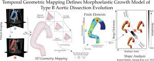
Temporal geometric mapping defines morphoelastic growth model of Type B aortic dissection evolution
The human aorta undergoes complex morphologic changes that mirror the evolution of disease. Finite element analysis (FEA) enables the prediction of aortic pathologic states, but the absence of a biomechanical understanding hinders the applicability of this computational tool. We incorporate geometric information from computed tomography angiography (CTA) imaging scans into FEA to predict a trajectory of future geometries for four aortic disease patients. Through defining a geometric correspondence between two patient scans separated in time, a patient-specific FEA model can recreate the deformation of the aorta between the two time points, showing that pathologic growth drives morphologic heterogeneity. FEA-derived trajectories in a shape-size geometric feature space, which plots the variance of the shape index versus the inverse square root of aortic surface area ( vs. ), quantitatively demonstrate an increase in . This represents a deviation from physiologic shape changes and parallels the true geometric progression of aortic disease patients.
期刊介绍:
Computers in Biology and Medicine is an international forum for sharing groundbreaking advancements in the use of computers in bioscience and medicine. This journal serves as a medium for communicating essential research, instruction, ideas, and information regarding the rapidly evolving field of computer applications in these domains. By encouraging the exchange of knowledge, we aim to facilitate progress and innovation in the utilization of computers in biology and medicine.


 分享
分享
 求助内容:
求助内容: 应助结果提醒方式:
 扫码关注我们
