Chen Liang , Yexuan Guo , Rui Xue Zhang , Hong Yan
{"title":"用于眼部多途径给药和用药的电纺聚乙二醇化聚(乳酸-共聚-乙醇酸)纤维植入物的微管和高孔隙率设计。","authors":"Chen Liang , Yexuan Guo , Rui Xue Zhang , Hong Yan","doi":"10.1016/j.ijpharm.2024.124751","DOIUrl":null,"url":null,"abstract":"<div><div>Electrospun fibers have been gaining popularity in ocular drug delivery and cellular therapies. However, most of electrospun fibers are planar-shape membrane with large dimension relative to intraocular space, making difficult to use as therapeutic implants. Herein, fibrous microtubes with a hollow center were fabricated by electrospinning using linear diblock mPEG<sub>2000</sub>-PLGA. Uniform microfibers with 0.809 μm diameter was tailored using Box-Behnken Design model for electrospinning process optimization. The microtubes were 1 mm long with a 0.386 mm diameter. Their suitability for intraocular administration was demonstrated by both injection <em>via</em> a 22-gauge needle and implant <em>via</em> integration of intraocular lens into the vitreous or anterior chamber of eyes, respectively. Electrospun mPEG<sub>2000</sub>-PLGA had higher porosity, smaller specific surface area, and smaller water contact angle, than that of PLGA. Macroscopically, mPEG<sub>2000</sub>-PLGA microfibers can maintain overall geometry upon exposure to aqueous buffer for 12 h while having high water uptake and exhibited good elasticity. Hydrolysis with 90 % polymeric degradation in 10.5 weeks underlied sustained slow release of anti-inflammatory drug dexamethasone. PEGylation of PLGA imparted preferential cell adhesion with markedly higher growth of human retinal epithelial cells than lens epithelial ones. This study highlights the potential utility of implantable electrospun PLGA-based microtubes for multiple intraocular delivery routes.</div></div>","PeriodicalId":14187,"journal":{"name":"International Journal of Pharmaceutics","volume":"665 ","pages":"Article 124751"},"PeriodicalIF":5.2000,"publicationDate":"2024-11-15","publicationTypes":"Journal Article","fieldsOfStudy":null,"isOpenAccess":false,"openAccessPdf":"","citationCount":"0","resultStr":"{\"title\":\"Microtubular and high porosity design of electrospun PEGylated poly (lactic-co-glycolic acid) fibrous implant for ocular multi-route administration and medication\",\"authors\":\"Chen Liang , Yexuan Guo , Rui Xue Zhang , Hong Yan\",\"doi\":\"10.1016/j.ijpharm.2024.124751\",\"DOIUrl\":null,\"url\":null,\"abstract\":\"<div><div>Electrospun fibers have been gaining popularity in ocular drug delivery and cellular therapies. However, most of electrospun fibers are planar-shape membrane with large dimension relative to intraocular space, making difficult to use as therapeutic implants. Herein, fibrous microtubes with a hollow center were fabricated by electrospinning using linear diblock mPEG<sub>2000</sub>-PLGA. Uniform microfibers with 0.809 μm diameter was tailored using Box-Behnken Design model for electrospinning process optimization. The microtubes were 1 mm long with a 0.386 mm diameter. Their suitability for intraocular administration was demonstrated by both injection <em>via</em> a 22-gauge needle and implant <em>via</em> integration of intraocular lens into the vitreous or anterior chamber of eyes, respectively. Electrospun mPEG<sub>2000</sub>-PLGA had higher porosity, smaller specific surface area, and smaller water contact angle, than that of PLGA. Macroscopically, mPEG<sub>2000</sub>-PLGA microfibers can maintain overall geometry upon exposure to aqueous buffer for 12 h while having high water uptake and exhibited good elasticity. Hydrolysis with 90 % polymeric degradation in 10.5 weeks underlied sustained slow release of anti-inflammatory drug dexamethasone. PEGylation of PLGA imparted preferential cell adhesion with markedly higher growth of human retinal epithelial cells than lens epithelial ones. This study highlights the potential utility of implantable electrospun PLGA-based microtubes for multiple intraocular delivery routes.</div></div>\",\"PeriodicalId\":14187,\"journal\":{\"name\":\"International Journal of Pharmaceutics\",\"volume\":\"665 \",\"pages\":\"Article 124751\"},\"PeriodicalIF\":5.2000,\"publicationDate\":\"2024-11-15\",\"publicationTypes\":\"Journal Article\",\"fieldsOfStudy\":null,\"isOpenAccess\":false,\"openAccessPdf\":\"\",\"citationCount\":\"0\",\"resultStr\":null,\"platform\":\"Semanticscholar\",\"paperid\":null,\"PeriodicalName\":\"International Journal of Pharmaceutics\",\"FirstCategoryId\":\"3\",\"ListUrlMain\":\"https://www.sciencedirect.com/science/article/pii/S0378517324009852\",\"RegionNum\":2,\"RegionCategory\":\"医学\",\"ArticlePicture\":[],\"TitleCN\":null,\"AbstractTextCN\":null,\"PMCID\":null,\"EPubDate\":\"2024/9/24 0:00:00\",\"PubModel\":\"Epub\",\"JCR\":\"Q1\",\"JCRName\":\"PHARMACOLOGY & PHARMACY\",\"Score\":null,\"Total\":0}","platform":"Semanticscholar","paperid":null,"PeriodicalName":"International Journal of Pharmaceutics","FirstCategoryId":"3","ListUrlMain":"https://www.sciencedirect.com/science/article/pii/S0378517324009852","RegionNum":2,"RegionCategory":"医学","ArticlePicture":[],"TitleCN":null,"AbstractTextCN":null,"PMCID":null,"EPubDate":"2024/9/24 0:00:00","PubModel":"Epub","JCR":"Q1","JCRName":"PHARMACOLOGY & PHARMACY","Score":null,"Total":0}
Microtubular and high porosity design of electrospun PEGylated poly (lactic-co-glycolic acid) fibrous implant for ocular multi-route administration and medication
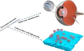
Electrospun fibers have been gaining popularity in ocular drug delivery and cellular therapies. However, most of electrospun fibers are planar-shape membrane with large dimension relative to intraocular space, making difficult to use as therapeutic implants. Herein, fibrous microtubes with a hollow center were fabricated by electrospinning using linear diblock mPEG2000-PLGA. Uniform microfibers with 0.809 μm diameter was tailored using Box-Behnken Design model for electrospinning process optimization. The microtubes were 1 mm long with a 0.386 mm diameter. Their suitability for intraocular administration was demonstrated by both injection via a 22-gauge needle and implant via integration of intraocular lens into the vitreous or anterior chamber of eyes, respectively. Electrospun mPEG2000-PLGA had higher porosity, smaller specific surface area, and smaller water contact angle, than that of PLGA. Macroscopically, mPEG2000-PLGA microfibers can maintain overall geometry upon exposure to aqueous buffer for 12 h while having high water uptake and exhibited good elasticity. Hydrolysis with 90 % polymeric degradation in 10.5 weeks underlied sustained slow release of anti-inflammatory drug dexamethasone. PEGylation of PLGA imparted preferential cell adhesion with markedly higher growth of human retinal epithelial cells than lens epithelial ones. This study highlights the potential utility of implantable electrospun PLGA-based microtubes for multiple intraocular delivery routes.
期刊介绍:
The International Journal of Pharmaceutics is the third most cited journal in the "Pharmacy & Pharmacology" category out of 366 journals, being the true home for pharmaceutical scientists concerned with the physical, chemical and biological properties of devices and delivery systems for drugs, vaccines and biologicals, including their design, manufacture and evaluation. This includes evaluation of the properties of drugs, excipients such as surfactants and polymers and novel materials. The journal has special sections on pharmaceutical nanotechnology and personalized medicines, and publishes research papers, reviews, commentaries and letters to the editor as well as special issues.


 分享
分享
 求助内容:
求助内容: 应助结果提醒方式:
 扫码关注我们
