Lukasz Stopa, Angelina Papeczyc, Zygmunt Stopa, Kamil Abed
{"title":"使用三维打印模型规划一名孤立性眶底骨折患者的手术治疗:病例报告。","authors":"Lukasz Stopa, Angelina Papeczyc, Zygmunt Stopa, Kamil Abed","doi":"10.21037/acr-24-73","DOIUrl":null,"url":null,"abstract":"<p><strong>Background: </strong>Orbital floor fractures typically manifest as eyeball mobility disorders with double vision (diplopia), enophthalmia, and infraorbital paresis. Surgical treatment of these fractures involves orbital floor reconstruction. The procedure involves freeing the trapped tissues from the lumen of the maxillary sinus and rebuilding the orbital floor. Technological progress in the field of three-dimensional (3D) printing allows physical prototyping of the implants to be used during the procedure.</p><p><strong>Case description: </strong>A 43-year-old female patient presented to the hospital with diplopia, which first occurred after a fall from own height. Examinations, including a computed tomography (CT) confirmed the diagnosis of an orbital floor fracture. 3D printing was used to plan the surgical treatment of the patient. Based on preoperative CT, a 1:1 scale model was prepared by means of 3D printing to demonstrate the fractured orbital area. It was later used to pre-cut a Codubix prosthesis, which was subsequently used to reconstruct the fractured bone. The patient's postoperative course was uneventful. Instant improvement in diplopia was noted. A CT scan was performed on the 3<sup>rd</sup> day after surgery. No herniation into the maxillary sinus was observed.</p><p><strong>Conclusions: </strong>3D printing seems to be a useful method that allows more thorough preparation for the surgery and also could potentially shorten its duration.</p>","PeriodicalId":29752,"journal":{"name":"AME Case Reports","volume":"8 ","pages":"110"},"PeriodicalIF":0.7000,"publicationDate":"2024-09-10","publicationTypes":"Journal Article","fieldsOfStudy":null,"isOpenAccess":false,"openAccessPdf":"https://www.ncbi.nlm.nih.gov/pmc/articles/PMC11459389/pdf/","citationCount":"0","resultStr":"{\"title\":\"Use of 3D-printed model to plan the surgical management of a patient with isolated orbital floor fracture: a case report.\",\"authors\":\"Lukasz Stopa, Angelina Papeczyc, Zygmunt Stopa, Kamil Abed\",\"doi\":\"10.21037/acr-24-73\",\"DOIUrl\":null,\"url\":null,\"abstract\":\"<p><strong>Background: </strong>Orbital floor fractures typically manifest as eyeball mobility disorders with double vision (diplopia), enophthalmia, and infraorbital paresis. Surgical treatment of these fractures involves orbital floor reconstruction. The procedure involves freeing the trapped tissues from the lumen of the maxillary sinus and rebuilding the orbital floor. Technological progress in the field of three-dimensional (3D) printing allows physical prototyping of the implants to be used during the procedure.</p><p><strong>Case description: </strong>A 43-year-old female patient presented to the hospital with diplopia, which first occurred after a fall from own height. Examinations, including a computed tomography (CT) confirmed the diagnosis of an orbital floor fracture. 3D printing was used to plan the surgical treatment of the patient. Based on preoperative CT, a 1:1 scale model was prepared by means of 3D printing to demonstrate the fractured orbital area. It was later used to pre-cut a Codubix prosthesis, which was subsequently used to reconstruct the fractured bone. The patient's postoperative course was uneventful. Instant improvement in diplopia was noted. A CT scan was performed on the 3<sup>rd</sup> day after surgery. No herniation into the maxillary sinus was observed.</p><p><strong>Conclusions: </strong>3D printing seems to be a useful method that allows more thorough preparation for the surgery and also could potentially shorten its duration.</p>\",\"PeriodicalId\":29752,\"journal\":{\"name\":\"AME Case Reports\",\"volume\":\"8 \",\"pages\":\"110\"},\"PeriodicalIF\":0.7000,\"publicationDate\":\"2024-09-10\",\"publicationTypes\":\"Journal Article\",\"fieldsOfStudy\":null,\"isOpenAccess\":false,\"openAccessPdf\":\"https://www.ncbi.nlm.nih.gov/pmc/articles/PMC11459389/pdf/\",\"citationCount\":\"0\",\"resultStr\":null,\"platform\":\"Semanticscholar\",\"paperid\":null,\"PeriodicalName\":\"AME Case Reports\",\"FirstCategoryId\":\"1085\",\"ListUrlMain\":\"https://doi.org/10.21037/acr-24-73\",\"RegionNum\":0,\"RegionCategory\":null,\"ArticlePicture\":[],\"TitleCN\":null,\"AbstractTextCN\":null,\"PMCID\":null,\"EPubDate\":\"2024/1/1 0:00:00\",\"PubModel\":\"eCollection\",\"JCR\":\"Q3\",\"JCRName\":\"MEDICINE, GENERAL & INTERNAL\",\"Score\":null,\"Total\":0}","platform":"Semanticscholar","paperid":null,"PeriodicalName":"AME Case Reports","FirstCategoryId":"1085","ListUrlMain":"https://doi.org/10.21037/acr-24-73","RegionNum":0,"RegionCategory":null,"ArticlePicture":[],"TitleCN":null,"AbstractTextCN":null,"PMCID":null,"EPubDate":"2024/1/1 0:00:00","PubModel":"eCollection","JCR":"Q3","JCRName":"MEDICINE, GENERAL & INTERNAL","Score":null,"Total":0}
引用次数: 0
摘要
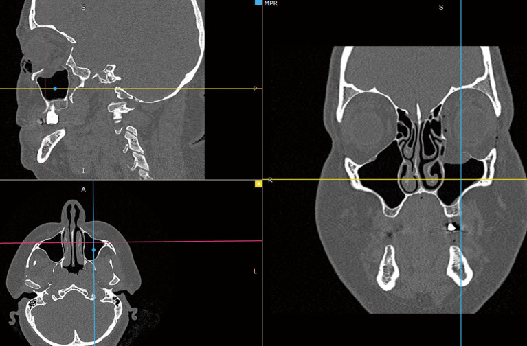
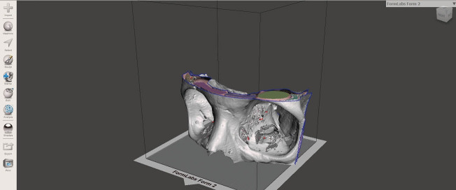
背景:眶底骨折通常表现为眼球活动障碍,伴有复视(复视)、眼球震颤和眶下瘫痪。这些骨折的手术治疗包括眶底重建。手术过程包括从上颌窦腔内释放被困组织,并重建眶底。三维(3D)打印领域的技术进步允许在手术过程中使用植入物的物理原型:一名 43 岁的女性患者因复视到医院就诊。包括计算机断层扫描(CT)在内的检查确诊为眶底骨折。患者的手术治疗计划采用了 3D 打印技术。根据术前 CT,通过 3D 打印技术制作了一个 1:1 比例的模型,以显示骨折的眼眶区域。随后,利用该模型预先切割了一个 Codubix 假体,用于重建骨折的骨头。患者术后恢复顺利。复视情况立即得到改善。术后第三天进行了 CT 扫描。结论:3D 打印似乎是一种有用的方法:3D打印似乎是一种有用的方法,可以为手术做更充分的准备,并有可能缩短手术时间。
Use of 3D-printed model to plan the surgical management of a patient with isolated orbital floor fracture: a case report.
Background: Orbital floor fractures typically manifest as eyeball mobility disorders with double vision (diplopia), enophthalmia, and infraorbital paresis. Surgical treatment of these fractures involves orbital floor reconstruction. The procedure involves freeing the trapped tissues from the lumen of the maxillary sinus and rebuilding the orbital floor. Technological progress in the field of three-dimensional (3D) printing allows physical prototyping of the implants to be used during the procedure.
Case description: A 43-year-old female patient presented to the hospital with diplopia, which first occurred after a fall from own height. Examinations, including a computed tomography (CT) confirmed the diagnosis of an orbital floor fracture. 3D printing was used to plan the surgical treatment of the patient. Based on preoperative CT, a 1:1 scale model was prepared by means of 3D printing to demonstrate the fractured orbital area. It was later used to pre-cut a Codubix prosthesis, which was subsequently used to reconstruct the fractured bone. The patient's postoperative course was uneventful. Instant improvement in diplopia was noted. A CT scan was performed on the 3rd day after surgery. No herniation into the maxillary sinus was observed.
Conclusions: 3D printing seems to be a useful method that allows more thorough preparation for the surgery and also could potentially shorten its duration.


 分享
分享

 求助内容:
求助内容: 应助结果提醒方式:
 扫码关注我们
