{"title":"对 \"发现新型组蛋白去乙酰化酶 6 (HDAC6) 抑制剂,增强黑色素瘤中抗 PD-L1 免疫疗法的抗肿瘤免疫力 \"的更正","authors":"Xiaopeng Peng, Ling Li, Jingxuan Chen, Yichang Ren, Jin Liu, Ziwen Yu, Hao Cao, Jianjun Chen","doi":"10.1021/acs.jmedchem.4c02486","DOIUrl":null,"url":null,"abstract":"In our study to investigate the impact of two newly synthesized compounds (XP5 and XP19) alongside a positive control drug (CAY10603) on the expression levels of various target proteins─specifically, AC-α-Tubulin, α-Tubulin, SMC3, Ac-H3, H3, Ac-SMC3, and apoptosis-related antibodies (Ac-Ku70, Ku70, Bcl-2, Bax)─we conducted a series of experiments on different tumor cell lines, including Jurkat-T cells and B16-F10 cells. This comprehensive analysis yielded a substantial amount of experimental data and protein bands. Regrettably, during the assembly of Figure 7 and Figure 8 in the original manuscript, we inadvertently used a wrong image of Ac-Ku70 and Ku70 in Figure 7A and of Ac-SMC3, SMC3, and Ac-H3 in Figure 8E,F. Upon recent and careful review of the original manuscript, we identified this error and have replaced the wrong image with the correct one in the corrected Figure 7 and Figure 8. This corrected result still demonstrates that XP5 and XP19 can upregulate the expression levels of acetylated Ku70 significantly and dose-dependently but showed no effect on the Ku70 levels and had little effect on the levels of acetyl-SMC3 and acetyl-H3, and these corrections do not change the scientific conclusions of the article in any way. Figure 7. Analysis of acetylated Ku70, Ku70, Bcl-2, and Bax by Western blot. Jurkat T cells were treated with dimethyl sulfoxide (DMSO) or XP5, and XP19 for 6 h. The levels of acetylated Ku70, Ku70, Bcl-2, and Bax were examined by Western blot (A). Quantitative analysis of the levels of acetylated Ku70 (B), Bcl-2 (C), and Bax (D). All data are representative of three independent experiments and shown as mean ± SD. ***<i>p</i> < 0.001, **<i>p</i> < 0.01, *<i>p</i> < 0.05 compared with the control group, <sup>###</sup><i>p</i> < 0.001. One-way ANOVA for the above analysis, Dunnett test. Figure 8. Analysis of acetylated α-tubulin/acetylated-SMC3 and acetylated-H3 by Western blot. The levels of acetyl-α-tubulin (Ac-α-Tub) and α-tubulin in B16-F10 cells (A) and Jurkat T cells (B) treated with DMSO or XP5, XP19, and CAY10603 for 6 h. Quantitative analysis of the level of acetyl-α-tubulin (Ac-α-Tub) by Western blotting assay obtained in B16-F10 cells (C) and Jurkat T cells (D). The levels of acetylated-SMC3, total SMC3, acetylated-H3, and total H3 in B16-F10 cells (E) and Jurkat T cells (F) treated with DMSO or XP5, XP19, and CAY10603 for 6 h. All data are representative of three independent experiments and shown as mean ± SD. ***<i>p</i> < 0.001, **<i>p</i> < 0.01 compared with the control group, <sup>###</sup><i>p</i> < 0.001, <sup>&</sup><i>p</i> < 0.05, <sup>&&</sup><i>p</i> < 0.01. One-way ANOVA for the above analysis, Dunnett test. We have now corrected the original Figure 7 and Figure 8, and all authors have agreed to the changes. We apologize for any confusion this may have caused and appreciate your understanding as we make these necessary corrections to ensure the integrity of our research findings. This article has not yet been cited by other publications.","PeriodicalId":46,"journal":{"name":"Journal of Medicinal Chemistry","volume":"233 1","pages":""},"PeriodicalIF":7.3000,"publicationDate":"2024-10-19","publicationTypes":"Journal Article","fieldsOfStudy":null,"isOpenAccess":false,"openAccessPdf":"","citationCount":"0","resultStr":"{\"title\":\"Correction to “Discovery of Novel Histone Deacetylase 6 (HDAC6) Inhibitors with Enhanced Antitumor Immunity of Anti-PD-L1 Immunotherapy in Melanoma”\",\"authors\":\"Xiaopeng Peng, Ling Li, Jingxuan Chen, Yichang Ren, Jin Liu, Ziwen Yu, Hao Cao, Jianjun Chen\",\"doi\":\"10.1021/acs.jmedchem.4c02486\",\"DOIUrl\":null,\"url\":null,\"abstract\":\"In our study to investigate the impact of two newly synthesized compounds (XP5 and XP19) alongside a positive control drug (CAY10603) on the expression levels of various target proteins─specifically, AC-α-Tubulin, α-Tubulin, SMC3, Ac-H3, H3, Ac-SMC3, and apoptosis-related antibodies (Ac-Ku70, Ku70, Bcl-2, Bax)─we conducted a series of experiments on different tumor cell lines, including Jurkat-T cells and B16-F10 cells. This comprehensive analysis yielded a substantial amount of experimental data and protein bands. Regrettably, during the assembly of Figure 7 and Figure 8 in the original manuscript, we inadvertently used a wrong image of Ac-Ku70 and Ku70 in Figure 7A and of Ac-SMC3, SMC3, and Ac-H3 in Figure 8E,F. Upon recent and careful review of the original manuscript, we identified this error and have replaced the wrong image with the correct one in the corrected Figure 7 and Figure 8. This corrected result still demonstrates that XP5 and XP19 can upregulate the expression levels of acetylated Ku70 significantly and dose-dependently but showed no effect on the Ku70 levels and had little effect on the levels of acetyl-SMC3 and acetyl-H3, and these corrections do not change the scientific conclusions of the article in any way. Figure 7. Analysis of acetylated Ku70, Ku70, Bcl-2, and Bax by Western blot. Jurkat T cells were treated with dimethyl sulfoxide (DMSO) or XP5, and XP19 for 6 h. The levels of acetylated Ku70, Ku70, Bcl-2, and Bax were examined by Western blot (A). Quantitative analysis of the levels of acetylated Ku70 (B), Bcl-2 (C), and Bax (D). All data are representative of three independent experiments and shown as mean ± SD. ***<i>p</i> < 0.001, **<i>p</i> < 0.01, *<i>p</i> < 0.05 compared with the control group, <sup>###</sup><i>p</i> < 0.001. One-way ANOVA for the above analysis, Dunnett test. Figure 8. Analysis of acetylated α-tubulin/acetylated-SMC3 and acetylated-H3 by Western blot. The levels of acetyl-α-tubulin (Ac-α-Tub) and α-tubulin in B16-F10 cells (A) and Jurkat T cells (B) treated with DMSO or XP5, XP19, and CAY10603 for 6 h. Quantitative analysis of the level of acetyl-α-tubulin (Ac-α-Tub) by Western blotting assay obtained in B16-F10 cells (C) and Jurkat T cells (D). The levels of acetylated-SMC3, total SMC3, acetylated-H3, and total H3 in B16-F10 cells (E) and Jurkat T cells (F) treated with DMSO or XP5, XP19, and CAY10603 for 6 h. All data are representative of three independent experiments and shown as mean ± SD. ***<i>p</i> < 0.001, **<i>p</i> < 0.01 compared with the control group, <sup>###</sup><i>p</i> < 0.001, <sup>&</sup><i>p</i> < 0.05, <sup>&&</sup><i>p</i> < 0.01. One-way ANOVA for the above analysis, Dunnett test. We have now corrected the original Figure 7 and Figure 8, and all authors have agreed to the changes. We apologize for any confusion this may have caused and appreciate your understanding as we make these necessary corrections to ensure the integrity of our research findings. This article has not yet been cited by other publications.\",\"PeriodicalId\":46,\"journal\":{\"name\":\"Journal of Medicinal Chemistry\",\"volume\":\"233 1\",\"pages\":\"\"},\"PeriodicalIF\":7.3000,\"publicationDate\":\"2024-10-19\",\"publicationTypes\":\"Journal Article\",\"fieldsOfStudy\":null,\"isOpenAccess\":false,\"openAccessPdf\":\"\",\"citationCount\":\"0\",\"resultStr\":null,\"platform\":\"Semanticscholar\",\"paperid\":null,\"PeriodicalName\":\"Journal of Medicinal Chemistry\",\"FirstCategoryId\":\"3\",\"ListUrlMain\":\"https://doi.org/10.1021/acs.jmedchem.4c02486\",\"RegionNum\":1,\"RegionCategory\":\"医学\",\"ArticlePicture\":[],\"TitleCN\":null,\"AbstractTextCN\":null,\"PMCID\":null,\"EPubDate\":\"\",\"PubModel\":\"\",\"JCR\":\"Q1\",\"JCRName\":\"CHEMISTRY, MEDICINAL\",\"Score\":null,\"Total\":0}","platform":"Semanticscholar","paperid":null,"PeriodicalName":"Journal of Medicinal Chemistry","FirstCategoryId":"3","ListUrlMain":"https://doi.org/10.1021/acs.jmedchem.4c02486","RegionNum":1,"RegionCategory":"医学","ArticlePicture":[],"TitleCN":null,"AbstractTextCN":null,"PMCID":null,"EPubDate":"","PubModel":"","JCR":"Q1","JCRName":"CHEMISTRY, MEDICINAL","Score":null,"Total":0}
Correction to “Discovery of Novel Histone Deacetylase 6 (HDAC6) Inhibitors with Enhanced Antitumor Immunity of Anti-PD-L1 Immunotherapy in Melanoma”
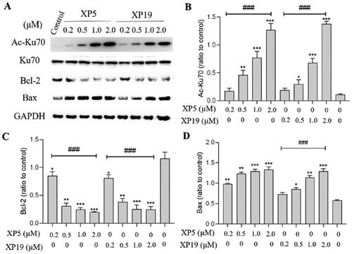
In our study to investigate the impact of two newly synthesized compounds (XP5 and XP19) alongside a positive control drug (CAY10603) on the expression levels of various target proteins─specifically, AC-α-Tubulin, α-Tubulin, SMC3, Ac-H3, H3, Ac-SMC3, and apoptosis-related antibodies (Ac-Ku70, Ku70, Bcl-2, Bax)─we conducted a series of experiments on different tumor cell lines, including Jurkat-T cells and B16-F10 cells. This comprehensive analysis yielded a substantial amount of experimental data and protein bands. Regrettably, during the assembly of Figure 7 and Figure 8 in the original manuscript, we inadvertently used a wrong image of Ac-Ku70 and Ku70 in Figure 7A and of Ac-SMC3, SMC3, and Ac-H3 in Figure 8E,F. Upon recent and careful review of the original manuscript, we identified this error and have replaced the wrong image with the correct one in the corrected Figure 7 and Figure 8. This corrected result still demonstrates that XP5 and XP19 can upregulate the expression levels of acetylated Ku70 significantly and dose-dependently but showed no effect on the Ku70 levels and had little effect on the levels of acetyl-SMC3 and acetyl-H3, and these corrections do not change the scientific conclusions of the article in any way. Figure 7. Analysis of acetylated Ku70, Ku70, Bcl-2, and Bax by Western blot. Jurkat T cells were treated with dimethyl sulfoxide (DMSO) or XP5, and XP19 for 6 h. The levels of acetylated Ku70, Ku70, Bcl-2, and Bax were examined by Western blot (A). Quantitative analysis of the levels of acetylated Ku70 (B), Bcl-2 (C), and Bax (D). All data are representative of three independent experiments and shown as mean ± SD. ***p < 0.001, **p < 0.01, *p < 0.05 compared with the control group, ###p < 0.001. One-way ANOVA for the above analysis, Dunnett test. Figure 8. Analysis of acetylated α-tubulin/acetylated-SMC3 and acetylated-H3 by Western blot. The levels of acetyl-α-tubulin (Ac-α-Tub) and α-tubulin in B16-F10 cells (A) and Jurkat T cells (B) treated with DMSO or XP5, XP19, and CAY10603 for 6 h. Quantitative analysis of the level of acetyl-α-tubulin (Ac-α-Tub) by Western blotting assay obtained in B16-F10 cells (C) and Jurkat T cells (D). The levels of acetylated-SMC3, total SMC3, acetylated-H3, and total H3 in B16-F10 cells (E) and Jurkat T cells (F) treated with DMSO or XP5, XP19, and CAY10603 for 6 h. All data are representative of three independent experiments and shown as mean ± SD. ***p < 0.001, **p < 0.01 compared with the control group, ###p < 0.001, &p < 0.05, &&p < 0.01. One-way ANOVA for the above analysis, Dunnett test. We have now corrected the original Figure 7 and Figure 8, and all authors have agreed to the changes. We apologize for any confusion this may have caused and appreciate your understanding as we make these necessary corrections to ensure the integrity of our research findings. This article has not yet been cited by other publications.
期刊介绍:
The Journal of Medicinal Chemistry is a prestigious biweekly peer-reviewed publication that focuses on the multifaceted field of medicinal chemistry. Since its inception in 1959 as the Journal of Medicinal and Pharmaceutical Chemistry, it has evolved to become a cornerstone in the dissemination of research findings related to the design, synthesis, and development of therapeutic agents.
The Journal of Medicinal Chemistry is recognized for its significant impact in the scientific community, as evidenced by its 2022 impact factor of 7.3. This metric reflects the journal's influence and the importance of its content in shaping the future of drug discovery and development. The journal serves as a vital resource for chemists, pharmacologists, and other researchers interested in the molecular mechanisms of drug action and the optimization of therapeutic compounds.


 分享
分享
 求助内容:
求助内容: 应助结果提醒方式:
 扫码关注我们
