{"title":"纳米颗粒封装的细胞铁(III):治疗肿瘤的 \"单刃剑\",具有微环境依赖性毒性","authors":"Yu Liu, Zhiyong Qian","doi":"10.1002/mog2.70001","DOIUrl":null,"url":null,"abstract":"<p>Recently, Li et al. published a study in <i>Science Advances</i> on the use of dual-responsive celastrol (CEL) materials for cancer treatment. The study took advantage of the high adenosine triphosphate (ATP) levels in the tumor microenvironment to modify the interior and surface of CEL, reducing its toxicity to normal tissues and maximizing the efficacy of its cancer treatment which contributed significantly to drug delivery for tumor therapy.<span><sup>1</sup></span></p><p>CEL is a drug with anticancer activity isolated from traditional Chinese medicinal herbs.<span><sup>2</sup></span> It inhibits cancer cell growth and survival signaling by binding to Cdc37 and Hsp90 reaction sites, causing disruption of the Hsp90-Cdc37 binding and folding of some of the client proteins.<span><sup>3</sup></span> However, its water stability, bioavailability, and targeting are undesirable, which limited its clinical application. There are currently two main approaches to promote CEL's therapeutic potential in clinic: 1. synthesizing derivatives by surface modification which mainly focuses on C-20 carboxylic acid functionality, alteration at the A ring, and C-6-modification at the B ring, and 2. adopting nanomedicine delivery systems, where liposomes, polymeric micelles, and nanoparticles (NPs) are commonly used. The second approach, however, requires strictly selected materials. Liposomes, with its ease of aggregation and instability and leakage when packaging drugs, and polymeric micelles, with potential toxicity, unclear drug delivery mechanisms, and poor quality/specification control, demand extra considerations in application. In this study an ATP/ROS dual-responsive CEL derivative was designed for tumor therapy by mainly taking advantage of the environment in which ATP levels inside and outside tumor cells are 10<sup>3</sup> to 10<sup>4</sup> times higher than that around normal tissues.<span><sup>4</sup></span> Surface modification of CEL using metal chelation was performed to address the toxicity to normal tissues, and NPs were selected for packaging to promote the release of CEL when it reaches the tumor site. Compared with liposomes and polymeric micelles, CEL-Fe with ATP/ROS dual-response has a well-defined structural study, low drug toxicity, high stability, and ultimately the ability to respond to the specificity of the tumor microenvironment for effective drug release and treatment which improves the biosafety and biocompatibility of the therapeutic drugs. Fe(III) was chosen to coordinately bond with oxygens on C-2 and C-3 in CEL to produce the less toxic CEL-Fe, which is then coated with polymer membrane consisting of thioketal-containing polyethylene glycol-grafted polymer and polyethylene enamine-modified F127 polymer to elevate its permeability and retention effects.<span><sup>5</sup></span> The high concentration of ATP in tumor tissues disrupts the coordination of Fe(III), restoring the pharmacological toxicity of CEL. In response to high level of ROS at tumor site, thioketal groups are degraded to facilitate drug release, and the oncology treatment is therefore initiated (Figure 1A).</p><p>The cell viability of cancer cells and normal cells were tested after treated with different groups: CEL, CEL-Fe, and CEL-Fe-ATP. The results verified the biosafety of CEL-Fe(III) chelate by showing its ATP-dependent cytotoxicity (Figures 1B,C). The mechanism of this property shift between CEL and CEL-Fe was explored by RNA sequencing analysis of A549 cells. PCA gene expression analysis showed that the CEL group and CEL-Fe+ATP group were clustered together, while separated from the CEL+Fe group. Moreover, the calculation of gene expression fold change and <i>p</i> Value plots revealed no significant gene expression difference between the CEL and CEL-Fe+ATP groups, while that of the CEL+Fe group varied. All above indicated the cytotoxicity weakening of the Fe(III) chelation and its restoration by ATP. The Kyoto Encyclopedia of Genes and Genomes (KEGG) analysis and Gene Set Enrichment Analysis showed a high enrichment of ER's protein processing, tumor necrosis factor signaling, and P53 signaling in CEL and CEL-Fe-ATP treatments, and a large degree of overlap of these pathways in CEL-Fe. This demonstrated that the functioning of CEL was related to the “life cycle” of protein, which was consistent with the mechanism of CEL antitumor therapy in the existing studies. The toxicity mechanism of CEL was further investigated where Hsp90/Cdc37 was chosen as the model for detection. Molecular docking and MD simulation revealed that CEL destroys the hydrogen bonds inside Hsp90-Cdc37 by occupying hydrophobic sites to cause protein degradation, while the number of hydrogen bonds destroyed by CEL-Fe is significantly smaller than that of CEL, which reduces its toxicity to the protein complex and cells. The data above provides a basis for a potential effective cancer therapy with this CEL derivative (Figures 1D,E).</p><p>In-vivo biosafety was examined by hema-toxylin and eosin (H&E) staining of tissue sections. Results showed that CEL and CEL NPs were significantly toxic to several vital tissues and organs, which did not occur in the CEL-Fe group. This demonstrated successful discrimination against tumor from normal tissues and organs brought by the metal chelation. Moreover, fluorescence imaging showed strong presence of CEL NPs at tumor sites in both cell-derived xenograft (CDX) and patient-derived xenograft (PDX) tumor models, demonstrating their superior biodispersibility and feasibility in future clinical trials.</p><p>Further studies were conducted by designing additional contrast groups of phosphate-buffered saline, CEL-Fe NPs, and NPs-treated mice with both CDX and PDX models to assess their antitumor ability. After the prescribed cycle, mice treated by CEL-Fe NPs in both models had the smallest tumor volume and best tumor treatment effect. Both H&E staining and TUNEL assay showed that the CEL-Fe NPs group ranked top in terms of tumor necrosis.</p><p>In conclusion, this study successfully synthesized a nanomaterial with ATP/ROS dual-responsiveness targeting mainly the high ATP level of the tumor microenvironment. The external packaging material was destroyed by Fenton reaction in the high ATP/ROS environment, exposing the loaded therapeutic drug CEL-Fe which was converted into CEL with toxicity restored by the high ATP concentration, exerting its full effect at the tumor site. In this study, the CEL modification cleverly solves CEL's therapeutic problem of discrimination between normal tissues and cancer tissues to achieve the ultimate goal of precision therapy at the tumor sites. This type of design is also the one of the main ideas of current tumor therapy, which takes advantage of the differences between the tumor microenvironment and that of the normal tissues. Such differences include ATP levels, pH, oxygen content, and so forthetc. It is highly expected that researchers may make good use of the intrinsic properties of tumor microenvironment to continue to study and develop drugs or drug delivery systems with ideal effectiveness, efficiency, and targeting.</p><p><b>Yu Liu</b>: Conception; drafting of the manuscript. <b>Zhiyong Qian</b>: Supervision. Both authors have read and approved the final version of the article.</p><p>The authors declare no conflict of interest.</p><p>Not applicable.</p>","PeriodicalId":100902,"journal":{"name":"MedComm – Oncology","volume":"3 4","pages":""},"PeriodicalIF":2.2000,"publicationDate":"2024-10-28","publicationTypes":"Journal Article","fieldsOfStudy":null,"isOpenAccess":false,"openAccessPdf":"https://onlinelibrary.wiley.com/doi/epdf/10.1002/mog2.70001","citationCount":"0","resultStr":"{\"title\":\"Nanoparticle-encapsuled celastrol-Fe(III): “Single-edged sword” for tumor therapy featuring microenvironment-dependent toxicity\",\"authors\":\"Yu Liu, Zhiyong Qian\",\"doi\":\"10.1002/mog2.70001\",\"DOIUrl\":null,\"url\":null,\"abstract\":\"<p>Recently, Li et al. published a study in <i>Science Advances</i> on the use of dual-responsive celastrol (CEL) materials for cancer treatment. The study took advantage of the high adenosine triphosphate (ATP) levels in the tumor microenvironment to modify the interior and surface of CEL, reducing its toxicity to normal tissues and maximizing the efficacy of its cancer treatment which contributed significantly to drug delivery for tumor therapy.<span><sup>1</sup></span></p><p>CEL is a drug with anticancer activity isolated from traditional Chinese medicinal herbs.<span><sup>2</sup></span> It inhibits cancer cell growth and survival signaling by binding to Cdc37 and Hsp90 reaction sites, causing disruption of the Hsp90-Cdc37 binding and folding of some of the client proteins.<span><sup>3</sup></span> However, its water stability, bioavailability, and targeting are undesirable, which limited its clinical application. There are currently two main approaches to promote CEL's therapeutic potential in clinic: 1. synthesizing derivatives by surface modification which mainly focuses on C-20 carboxylic acid functionality, alteration at the A ring, and C-6-modification at the B ring, and 2. adopting nanomedicine delivery systems, where liposomes, polymeric micelles, and nanoparticles (NPs) are commonly used. The second approach, however, requires strictly selected materials. Liposomes, with its ease of aggregation and instability and leakage when packaging drugs, and polymeric micelles, with potential toxicity, unclear drug delivery mechanisms, and poor quality/specification control, demand extra considerations in application. In this study an ATP/ROS dual-responsive CEL derivative was designed for tumor therapy by mainly taking advantage of the environment in which ATP levels inside and outside tumor cells are 10<sup>3</sup> to 10<sup>4</sup> times higher than that around normal tissues.<span><sup>4</sup></span> Surface modification of CEL using metal chelation was performed to address the toxicity to normal tissues, and NPs were selected for packaging to promote the release of CEL when it reaches the tumor site. Compared with liposomes and polymeric micelles, CEL-Fe with ATP/ROS dual-response has a well-defined structural study, low drug toxicity, high stability, and ultimately the ability to respond to the specificity of the tumor microenvironment for effective drug release and treatment which improves the biosafety and biocompatibility of the therapeutic drugs. Fe(III) was chosen to coordinately bond with oxygens on C-2 and C-3 in CEL to produce the less toxic CEL-Fe, which is then coated with polymer membrane consisting of thioketal-containing polyethylene glycol-grafted polymer and polyethylene enamine-modified F127 polymer to elevate its permeability and retention effects.<span><sup>5</sup></span> The high concentration of ATP in tumor tissues disrupts the coordination of Fe(III), restoring the pharmacological toxicity of CEL. In response to high level of ROS at tumor site, thioketal groups are degraded to facilitate drug release, and the oncology treatment is therefore initiated (Figure 1A).</p><p>The cell viability of cancer cells and normal cells were tested after treated with different groups: CEL, CEL-Fe, and CEL-Fe-ATP. The results verified the biosafety of CEL-Fe(III) chelate by showing its ATP-dependent cytotoxicity (Figures 1B,C). The mechanism of this property shift between CEL and CEL-Fe was explored by RNA sequencing analysis of A549 cells. PCA gene expression analysis showed that the CEL group and CEL-Fe+ATP group were clustered together, while separated from the CEL+Fe group. Moreover, the calculation of gene expression fold change and <i>p</i> Value plots revealed no significant gene expression difference between the CEL and CEL-Fe+ATP groups, while that of the CEL+Fe group varied. All above indicated the cytotoxicity weakening of the Fe(III) chelation and its restoration by ATP. The Kyoto Encyclopedia of Genes and Genomes (KEGG) analysis and Gene Set Enrichment Analysis showed a high enrichment of ER's protein processing, tumor necrosis factor signaling, and P53 signaling in CEL and CEL-Fe-ATP treatments, and a large degree of overlap of these pathways in CEL-Fe. This demonstrated that the functioning of CEL was related to the “life cycle” of protein, which was consistent with the mechanism of CEL antitumor therapy in the existing studies. The toxicity mechanism of CEL was further investigated where Hsp90/Cdc37 was chosen as the model for detection. Molecular docking and MD simulation revealed that CEL destroys the hydrogen bonds inside Hsp90-Cdc37 by occupying hydrophobic sites to cause protein degradation, while the number of hydrogen bonds destroyed by CEL-Fe is significantly smaller than that of CEL, which reduces its toxicity to the protein complex and cells. The data above provides a basis for a potential effective cancer therapy with this CEL derivative (Figures 1D,E).</p><p>In-vivo biosafety was examined by hema-toxylin and eosin (H&E) staining of tissue sections. Results showed that CEL and CEL NPs were significantly toxic to several vital tissues and organs, which did not occur in the CEL-Fe group. This demonstrated successful discrimination against tumor from normal tissues and organs brought by the metal chelation. Moreover, fluorescence imaging showed strong presence of CEL NPs at tumor sites in both cell-derived xenograft (CDX) and patient-derived xenograft (PDX) tumor models, demonstrating their superior biodispersibility and feasibility in future clinical trials.</p><p>Further studies were conducted by designing additional contrast groups of phosphate-buffered saline, CEL-Fe NPs, and NPs-treated mice with both CDX and PDX models to assess their antitumor ability. After the prescribed cycle, mice treated by CEL-Fe NPs in both models had the smallest tumor volume and best tumor treatment effect. Both H&E staining and TUNEL assay showed that the CEL-Fe NPs group ranked top in terms of tumor necrosis.</p><p>In conclusion, this study successfully synthesized a nanomaterial with ATP/ROS dual-responsiveness targeting mainly the high ATP level of the tumor microenvironment. The external packaging material was destroyed by Fenton reaction in the high ATP/ROS environment, exposing the loaded therapeutic drug CEL-Fe which was converted into CEL with toxicity restored by the high ATP concentration, exerting its full effect at the tumor site. In this study, the CEL modification cleverly solves CEL's therapeutic problem of discrimination between normal tissues and cancer tissues to achieve the ultimate goal of precision therapy at the tumor sites. This type of design is also the one of the main ideas of current tumor therapy, which takes advantage of the differences between the tumor microenvironment and that of the normal tissues. Such differences include ATP levels, pH, oxygen content, and so forthetc. It is highly expected that researchers may make good use of the intrinsic properties of tumor microenvironment to continue to study and develop drugs or drug delivery systems with ideal effectiveness, efficiency, and targeting.</p><p><b>Yu Liu</b>: Conception; drafting of the manuscript. <b>Zhiyong Qian</b>: Supervision. Both authors have read and approved the final version of the article.</p><p>The authors declare no conflict of interest.</p><p>Not applicable.</p>\",\"PeriodicalId\":100902,\"journal\":{\"name\":\"MedComm – Oncology\",\"volume\":\"3 4\",\"pages\":\"\"},\"PeriodicalIF\":2.2000,\"publicationDate\":\"2024-10-28\",\"publicationTypes\":\"Journal Article\",\"fieldsOfStudy\":null,\"isOpenAccess\":false,\"openAccessPdf\":\"https://onlinelibrary.wiley.com/doi/epdf/10.1002/mog2.70001\",\"citationCount\":\"0\",\"resultStr\":null,\"platform\":\"Semanticscholar\",\"paperid\":null,\"PeriodicalName\":\"MedComm – Oncology\",\"FirstCategoryId\":\"1085\",\"ListUrlMain\":\"https://onlinelibrary.wiley.com/doi/10.1002/mog2.70001\",\"RegionNum\":0,\"RegionCategory\":null,\"ArticlePicture\":[],\"TitleCN\":null,\"AbstractTextCN\":null,\"PMCID\":null,\"EPubDate\":\"\",\"PubModel\":\"\",\"JCR\":\"\",\"JCRName\":\"\",\"Score\":null,\"Total\":0}","platform":"Semanticscholar","paperid":null,"PeriodicalName":"MedComm – Oncology","FirstCategoryId":"1085","ListUrlMain":"https://onlinelibrary.wiley.com/doi/10.1002/mog2.70001","RegionNum":0,"RegionCategory":null,"ArticlePicture":[],"TitleCN":null,"AbstractTextCN":null,"PMCID":null,"EPubDate":"","PubModel":"","JCR":"","JCRName":"","Score":null,"Total":0}
引用次数: 0
摘要
最近,Li 等人在《科学进展》(Science Advances)杂志上发表了一项关于使用双响应糜蛋白(CEL)材料治疗癌症的研究。该研究利用肿瘤微环境中的高三磷酸腺苷(ATP)水平,对 CEL 的内部和表面进行了改性,降低了其对正常组织的毒性,最大限度地提高了其癌症治疗效果,为肿瘤治疗的药物输送做出了重要贡献。2 它通过与 Cdc37 和 Hsp90 反应位点结合,破坏 Hsp90-Cdc37 的结合和部分客户蛋白的折叠,从而抑制癌细胞的生长和存活信号传导。目前,促进 CEL 临床治疗潜力的方法主要有两种:1. 通过表面改性合成衍生物,主要集中在 C-20 羧酸官能团、A 环的改变和 B 环的 C-6 改性;以及 2. 采用纳米药物传输系统,常用的有脂质体、聚合物胶束和纳米颗粒(NPs)。不过,第二种方法需要严格选择材料。脂质体在包装药物时容易聚集、不稳定和泄漏,而聚合物胶束则有潜在毒性、给药机制不明确、质量/规格控制不佳等问题,因此在应用时需要格外注意。本研究主要利用肿瘤细胞内外的 ATP 水平是正常组织周围的 103-104 倍这一环境,设计了一种 ATP/ROS 双响应 CEL 衍生物用于肿瘤治疗。与脂质体和高分子胶束相比,具有 ATP/ROS 双响应的 CEL-Fe 具有结构研究明确、药物毒性低、稳定性高的特点,最终能够响应肿瘤微环境的特异性,实现有效的药物释放和治疗,提高了治疗药物的生物安全性和生物相容性。选择铁(III)与 CEL 中 C-2 和 C-3 上的氧原子配位结合,生成毒性较低的 CEL-Fe,然后将其包覆在由含硫酮的聚乙二醇接枝聚合物和聚乙烯烯胺改性 F127 聚合物组成的聚合物膜上,以提高其渗透性和保留效果5。肿瘤组织中高浓度的 ATP 破坏了铁(III)的配位,从而恢复了 CEL 的药理毒性。在肿瘤部位高浓度的 ROS 作用下,硫酮基团被降解,促进了药物的释放,从而启动了肿瘤治疗(图 1A):经 CEL、CEL-Fe 和 CEL-Fe-ATP 处理后,对癌细胞和正常细胞的存活率进行了测试。结果表明,CEL-Fe(III)螯合物具有 ATP 依赖性细胞毒性,从而验证了其生物安全性(图 1B、C)。我们通过对 A549 细胞进行 RNA 测序分析,探索了 CEL 和 CEL-Fe 性质转变的机制。PCA 基因表达分析表明,CEL 组和 CEL-Fe+ATP 组聚集在一起,而与 CEL+Fe 组分开。此外,通过计算基因表达量折叠变化和 p 值图,发现 CEL 组和 CEL-Fe+ATP 组的基因表达量无显著差异,而 CEL+Fe 组的基因表达量有差异。这表明铁(III)螯合作用的细胞毒性减弱,而 ATP 作用的细胞毒性恢复。京都基因组百科全书(KEGG)分析和基因组富集分析表明,ER蛋白处理、肿瘤坏死因子信号转导和P53信号转导在CEL和CEL-Fe-ATP处理中富集程度较高,而在CEL-Fe处理中这些通路有很大程度的重叠。这表明CEL的功能与蛋白质的 "生命周期 "有关,这与现有研究中CEL抗肿瘤治疗的机制是一致的。研究人员进一步研究了CEL的毒性机制,并选择Hsp90/Cdc37作为检测模型。分子对接和 MD 模拟显示,CEL 通过占据疏水位点破坏 Hsp90-Cdc37 内部的氢键,导致蛋白质降解,而 CEL-Fe 破坏的氢键数量明显少于 CEL,从而降低了其对蛋白质复合物和细胞的毒性。上述数据为利用这种 CEL 衍生物进行有效的癌症治疗提供了依据(图 1D、E)。组织切片的hema-toxylin 和 eosin(H&E)染色检验了体内生物安全性。 结果表明,CEL 和 CEL NPs 对多个重要组织和器官有明显毒性,而 CEL-Fe 组则没有。这表明金属螯合作用成功地将肿瘤与正常组织器官区分开来。此外,荧光成像显示,在细胞衍生异种移植(CDX)和患者衍生异种移植(PDX)肿瘤模型中,CEL NPs 在肿瘤部位都有很强的存在性,这证明了其优越的生物分散性和在未来临床试验中的可行性。在规定周期后,CEL-Fe NPs 治疗的两种模型小鼠的肿瘤体积最小,肿瘤治疗效果最好。H&E染色和TUNEL检测均显示,CEL-Fe NPs组的肿瘤坏死率名列前茅。外部包装材料在高 ATP/ROS 环境中被 Fenton 反应破坏,暴露出负载的治疗药物 CEL-Fe,CEL-Fe 在高 ATP 浓度的作用下转化为 CEL,毒性得到恢复,在肿瘤部位充分发挥作用。在这项研究中,对 CEL 的改造巧妙地解决了 CEL 在治疗上区分正常组织和癌症组织的问题,实现了在肿瘤部位进行精准治疗的最终目标。这种设计也是目前肿瘤治疗的主要思路之一,即利用肿瘤微环境与正常组织微环境的差异。这种差异包括 ATP 水平、pH 值、氧含量等。希望研究人员能充分利用肿瘤微环境的内在特性,继续研究和开发具有理想疗效、效率和靶向性的药物或给药系统。钱志勇指导。作者声明无利益冲突。
Nanoparticle-encapsuled celastrol-Fe(III): “Single-edged sword” for tumor therapy featuring microenvironment-dependent toxicity
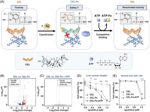
Recently, Li et al. published a study in Science Advances on the use of dual-responsive celastrol (CEL) materials for cancer treatment. The study took advantage of the high adenosine triphosphate (ATP) levels in the tumor microenvironment to modify the interior and surface of CEL, reducing its toxicity to normal tissues and maximizing the efficacy of its cancer treatment which contributed significantly to drug delivery for tumor therapy.1
CEL is a drug with anticancer activity isolated from traditional Chinese medicinal herbs.2 It inhibits cancer cell growth and survival signaling by binding to Cdc37 and Hsp90 reaction sites, causing disruption of the Hsp90-Cdc37 binding and folding of some of the client proteins.3 However, its water stability, bioavailability, and targeting are undesirable, which limited its clinical application. There are currently two main approaches to promote CEL's therapeutic potential in clinic: 1. synthesizing derivatives by surface modification which mainly focuses on C-20 carboxylic acid functionality, alteration at the A ring, and C-6-modification at the B ring, and 2. adopting nanomedicine delivery systems, where liposomes, polymeric micelles, and nanoparticles (NPs) are commonly used. The second approach, however, requires strictly selected materials. Liposomes, with its ease of aggregation and instability and leakage when packaging drugs, and polymeric micelles, with potential toxicity, unclear drug delivery mechanisms, and poor quality/specification control, demand extra considerations in application. In this study an ATP/ROS dual-responsive CEL derivative was designed for tumor therapy by mainly taking advantage of the environment in which ATP levels inside and outside tumor cells are 103 to 104 times higher than that around normal tissues.4 Surface modification of CEL using metal chelation was performed to address the toxicity to normal tissues, and NPs were selected for packaging to promote the release of CEL when it reaches the tumor site. Compared with liposomes and polymeric micelles, CEL-Fe with ATP/ROS dual-response has a well-defined structural study, low drug toxicity, high stability, and ultimately the ability to respond to the specificity of the tumor microenvironment for effective drug release and treatment which improves the biosafety and biocompatibility of the therapeutic drugs. Fe(III) was chosen to coordinately bond with oxygens on C-2 and C-3 in CEL to produce the less toxic CEL-Fe, which is then coated with polymer membrane consisting of thioketal-containing polyethylene glycol-grafted polymer and polyethylene enamine-modified F127 polymer to elevate its permeability and retention effects.5 The high concentration of ATP in tumor tissues disrupts the coordination of Fe(III), restoring the pharmacological toxicity of CEL. In response to high level of ROS at tumor site, thioketal groups are degraded to facilitate drug release, and the oncology treatment is therefore initiated (Figure 1A).
The cell viability of cancer cells and normal cells were tested after treated with different groups: CEL, CEL-Fe, and CEL-Fe-ATP. The results verified the biosafety of CEL-Fe(III) chelate by showing its ATP-dependent cytotoxicity (Figures 1B,C). The mechanism of this property shift between CEL and CEL-Fe was explored by RNA sequencing analysis of A549 cells. PCA gene expression analysis showed that the CEL group and CEL-Fe+ATP group were clustered together, while separated from the CEL+Fe group. Moreover, the calculation of gene expression fold change and p Value plots revealed no significant gene expression difference between the CEL and CEL-Fe+ATP groups, while that of the CEL+Fe group varied. All above indicated the cytotoxicity weakening of the Fe(III) chelation and its restoration by ATP. The Kyoto Encyclopedia of Genes and Genomes (KEGG) analysis and Gene Set Enrichment Analysis showed a high enrichment of ER's protein processing, tumor necrosis factor signaling, and P53 signaling in CEL and CEL-Fe-ATP treatments, and a large degree of overlap of these pathways in CEL-Fe. This demonstrated that the functioning of CEL was related to the “life cycle” of protein, which was consistent with the mechanism of CEL antitumor therapy in the existing studies. The toxicity mechanism of CEL was further investigated where Hsp90/Cdc37 was chosen as the model for detection. Molecular docking and MD simulation revealed that CEL destroys the hydrogen bonds inside Hsp90-Cdc37 by occupying hydrophobic sites to cause protein degradation, while the number of hydrogen bonds destroyed by CEL-Fe is significantly smaller than that of CEL, which reduces its toxicity to the protein complex and cells. The data above provides a basis for a potential effective cancer therapy with this CEL derivative (Figures 1D,E).
In-vivo biosafety was examined by hema-toxylin and eosin (H&E) staining of tissue sections. Results showed that CEL and CEL NPs were significantly toxic to several vital tissues and organs, which did not occur in the CEL-Fe group. This demonstrated successful discrimination against tumor from normal tissues and organs brought by the metal chelation. Moreover, fluorescence imaging showed strong presence of CEL NPs at tumor sites in both cell-derived xenograft (CDX) and patient-derived xenograft (PDX) tumor models, demonstrating their superior biodispersibility and feasibility in future clinical trials.
Further studies were conducted by designing additional contrast groups of phosphate-buffered saline, CEL-Fe NPs, and NPs-treated mice with both CDX and PDX models to assess their antitumor ability. After the prescribed cycle, mice treated by CEL-Fe NPs in both models had the smallest tumor volume and best tumor treatment effect. Both H&E staining and TUNEL assay showed that the CEL-Fe NPs group ranked top in terms of tumor necrosis.
In conclusion, this study successfully synthesized a nanomaterial with ATP/ROS dual-responsiveness targeting mainly the high ATP level of the tumor microenvironment. The external packaging material was destroyed by Fenton reaction in the high ATP/ROS environment, exposing the loaded therapeutic drug CEL-Fe which was converted into CEL with toxicity restored by the high ATP concentration, exerting its full effect at the tumor site. In this study, the CEL modification cleverly solves CEL's therapeutic problem of discrimination between normal tissues and cancer tissues to achieve the ultimate goal of precision therapy at the tumor sites. This type of design is also the one of the main ideas of current tumor therapy, which takes advantage of the differences between the tumor microenvironment and that of the normal tissues. Such differences include ATP levels, pH, oxygen content, and so forthetc. It is highly expected that researchers may make good use of the intrinsic properties of tumor microenvironment to continue to study and develop drugs or drug delivery systems with ideal effectiveness, efficiency, and targeting.
Yu Liu: Conception; drafting of the manuscript. Zhiyong Qian: Supervision. Both authors have read and approved the final version of the article.


 分享
分享

 求助内容:
求助内容: 应助结果提醒方式:
 扫码关注我们
