Mustafa Kayabasi, Seher Koksaldi, Ali Osman Saatci
{"title":"视网膜内高反射线:各种视网膜疾病的潜在生物标记物。","authors":"Mustafa Kayabasi, Seher Koksaldi, Ali Osman Saatci","doi":"10.51329/mehdiophthal1504","DOIUrl":null,"url":null,"abstract":"<p><strong>Background: </strong>The intraretinal hyperreflective line (IHL) is a novel posterior segment finding demonstrable using careful optical coherence tomography (OCT) examination. It likely indicates a reaction against photoreceptor, Muller cell, and/or retinal pigment epithelial damage. This study analyzed the spectral-domain OCT characteristics of IHLs to disclose their presence in various retinal conditions.</p><p><strong>Methods: </strong>A retrospective review of the charted and imaging records of participants with IHL was conducted at Dokuz Eylul University Department of Ophthalmology between January 2019 and August 2023. The inclusion criterion was the detection of an IHL on good-quality B-scan spectral-domain OCT. An IHL was defined as a vertical line extending from the ellipsoid zone band (or lower) through the outer nuclear layer to the internal limiting membrane in the central fovea. Associated retinal conditions were recorded as potential causative factors for the presence of IHL.</p><p><strong>Results: </strong>IHL was observed on spectral-domain OCT in 40 eyes of 38 participants with several retinal diseases assessment. Fourteen eyes (35%) underwent vitreoretinal surgery pre-IHL detection (12 were operated for full-thickness macular hole [FTMH], one for epiretinal membrane [ERM], and one for rhegmatogenous retinal detachment). In six eyes (15%) a microhole coexisted. Four eyes (10%) had a concurrent lamellar macular hole. The IHL preceded the occurrence of FTMH in three eyes (7.5%), and diabetic macular edema and type 2 idiopathic macular telangiectasia (MacTel-2) were present in three eyes (7.5%) each. The remaining conditions included vitreomacular traction (VMT), nonarteritic anterior ischemic optic neuropathy with central retinal artery occlusion, commotio retinae, exudative age-related macular degeneration, ERM, non-infectious idiopathic posterior uveitis, and Coats' disease, each affecting one eye (2.5%). Of the two participants with bilateral involvement, one was diagnosed with MacTel-2 and the other had IHL with VMT in the right eye that was detected post-vitreoretinal surgery for FTMH in the left eye.</p><p><strong>Conclusions: </strong>Although IHLs are mostly identified in eyes with vitreomacular surface diseases, clinicians may encounter IHLs in other types of retinal pathology. Further large-scale, multicenter, long-term studies on the presence of IHLs in OCT imaging are required to provide more substantial insight on this biomarker.</p>","PeriodicalId":36524,"journal":{"name":"Medical Hypothesis, Discovery, and Innovation in Ophthalmology","volume":"13 3","pages":"129-138"},"PeriodicalIF":0.0000,"publicationDate":"2024-10-14","publicationTypes":"Journal Article","fieldsOfStudy":null,"isOpenAccess":false,"openAccessPdf":"https://www.ncbi.nlm.nih.gov/pmc/articles/PMC11537236/pdf/","citationCount":"0","resultStr":"{\"title\":\"Intraretinal hyperreflective line: potential biomarker in various retinal disorders.\",\"authors\":\"Mustafa Kayabasi, Seher Koksaldi, Ali Osman Saatci\",\"doi\":\"10.51329/mehdiophthal1504\",\"DOIUrl\":null,\"url\":null,\"abstract\":\"<p><strong>Background: </strong>The intraretinal hyperreflective line (IHL) is a novel posterior segment finding demonstrable using careful optical coherence tomography (OCT) examination. It likely indicates a reaction against photoreceptor, Muller cell, and/or retinal pigment epithelial damage. This study analyzed the spectral-domain OCT characteristics of IHLs to disclose their presence in various retinal conditions.</p><p><strong>Methods: </strong>A retrospective review of the charted and imaging records of participants with IHL was conducted at Dokuz Eylul University Department of Ophthalmology between January 2019 and August 2023. The inclusion criterion was the detection of an IHL on good-quality B-scan spectral-domain OCT. An IHL was defined as a vertical line extending from the ellipsoid zone band (or lower) through the outer nuclear layer to the internal limiting membrane in the central fovea. Associated retinal conditions were recorded as potential causative factors for the presence of IHL.</p><p><strong>Results: </strong>IHL was observed on spectral-domain OCT in 40 eyes of 38 participants with several retinal diseases assessment. Fourteen eyes (35%) underwent vitreoretinal surgery pre-IHL detection (12 were operated for full-thickness macular hole [FTMH], one for epiretinal membrane [ERM], and one for rhegmatogenous retinal detachment). In six eyes (15%) a microhole coexisted. Four eyes (10%) had a concurrent lamellar macular hole. The IHL preceded the occurrence of FTMH in three eyes (7.5%), and diabetic macular edema and type 2 idiopathic macular telangiectasia (MacTel-2) were present in three eyes (7.5%) each. The remaining conditions included vitreomacular traction (VMT), nonarteritic anterior ischemic optic neuropathy with central retinal artery occlusion, commotio retinae, exudative age-related macular degeneration, ERM, non-infectious idiopathic posterior uveitis, and Coats' disease, each affecting one eye (2.5%). Of the two participants with bilateral involvement, one was diagnosed with MacTel-2 and the other had IHL with VMT in the right eye that was detected post-vitreoretinal surgery for FTMH in the left eye.</p><p><strong>Conclusions: </strong>Although IHLs are mostly identified in eyes with vitreomacular surface diseases, clinicians may encounter IHLs in other types of retinal pathology. Further large-scale, multicenter, long-term studies on the presence of IHLs in OCT imaging are required to provide more substantial insight on this biomarker.</p>\",\"PeriodicalId\":36524,\"journal\":{\"name\":\"Medical Hypothesis, Discovery, and Innovation in Ophthalmology\",\"volume\":\"13 3\",\"pages\":\"129-138\"},\"PeriodicalIF\":0.0000,\"publicationDate\":\"2024-10-14\",\"publicationTypes\":\"Journal Article\",\"fieldsOfStudy\":null,\"isOpenAccess\":false,\"openAccessPdf\":\"https://www.ncbi.nlm.nih.gov/pmc/articles/PMC11537236/pdf/\",\"citationCount\":\"0\",\"resultStr\":null,\"platform\":\"Semanticscholar\",\"paperid\":null,\"PeriodicalName\":\"Medical Hypothesis, Discovery, and Innovation in Ophthalmology\",\"FirstCategoryId\":\"1085\",\"ListUrlMain\":\"https://doi.org/10.51329/mehdiophthal1504\",\"RegionNum\":0,\"RegionCategory\":null,\"ArticlePicture\":[],\"TitleCN\":null,\"AbstractTextCN\":null,\"PMCID\":null,\"EPubDate\":\"2024/1/1 0:00:00\",\"PubModel\":\"eCollection\",\"JCR\":\"Q2\",\"JCRName\":\"Medicine\",\"Score\":null,\"Total\":0}","platform":"Semanticscholar","paperid":null,"PeriodicalName":"Medical Hypothesis, Discovery, and Innovation in Ophthalmology","FirstCategoryId":"1085","ListUrlMain":"https://doi.org/10.51329/mehdiophthal1504","RegionNum":0,"RegionCategory":null,"ArticlePicture":[],"TitleCN":null,"AbstractTextCN":null,"PMCID":null,"EPubDate":"2024/1/1 0:00:00","PubModel":"eCollection","JCR":"Q2","JCRName":"Medicine","Score":null,"Total":0}
引用次数: 0
摘要
背景:视网膜内高反光线(IHL)是一种新的后段发现,通过仔细的光学相干断层扫描(OCT)检查可以发现。它可能是对感光细胞、Muller 细胞和/或视网膜色素上皮损伤的一种反应。本研究分析了IHL的光谱域OCT特征,以揭示其在各种视网膜病症中的存在:方法:2019 年 1 月至 2023 年 8 月期间,Dokuz Eylul 大学眼科部对 IHL 患者的病历和成像记录进行了回顾性审查。纳入标准是在优质 B 扫描光谱域 OCT 上检测到 IHL。IHL被定义为一条从椭圆带(或更低)穿过核外层延伸至中央眼窝内缘膜的垂直线。相关视网膜条件被记录下来,作为出现 IHL 的潜在致病因素:结果:38 名参与者的 40 只眼睛在光谱域 OCT 上观察到 IHL,并对其进行了多种视网膜疾病评估。14只眼睛(35%)在检测到IHL之前接受了玻璃体视网膜手术(12只眼睛因全厚黄斑孔[FTMH]接受手术,1只眼睛因视网膜上膜[ERM]接受手术,1只眼睛因流变性视网膜脱离接受手术)。六只眼(15%)同时存在微孔。四只眼(10%)同时存在片状黄斑孔。有三只眼(7.5%)在发生 FTMH 之前出现了 IHL,有三只眼(7.5%)分别出现了糖尿病性黄斑水肿和 2 型特发性黄斑毛细血管扩张症(MacTel-2)。其余病症包括玻璃体黄斑牵引(VMT)、视网膜中央动脉闭塞性非动脉炎性前部缺血性视神经病变、视网膜皲裂、渗出性老年性黄斑变性、ERM、非感染性特发性后葡萄膜炎和 Coats 病,各影响一只眼(2.5%)。在两名双侧受累的参与者中,一人被诊断为MacTel-2,另一人右眼患有IHL并伴有VMT,是在左眼因FTMH进行玻璃体视网膜手术后被发现的:结论:虽然IHL多见于玻璃体视网膜表面疾病,但临床医生也可能在其他类型的视网膜病变中发现IHL。需要进一步开展大规模、多中心、长期的 OCT 成像中 IHLs 存在情况的研究,以便对这一生物标志物有更深入的了解。
Intraretinal hyperreflective line: potential biomarker in various retinal disorders.
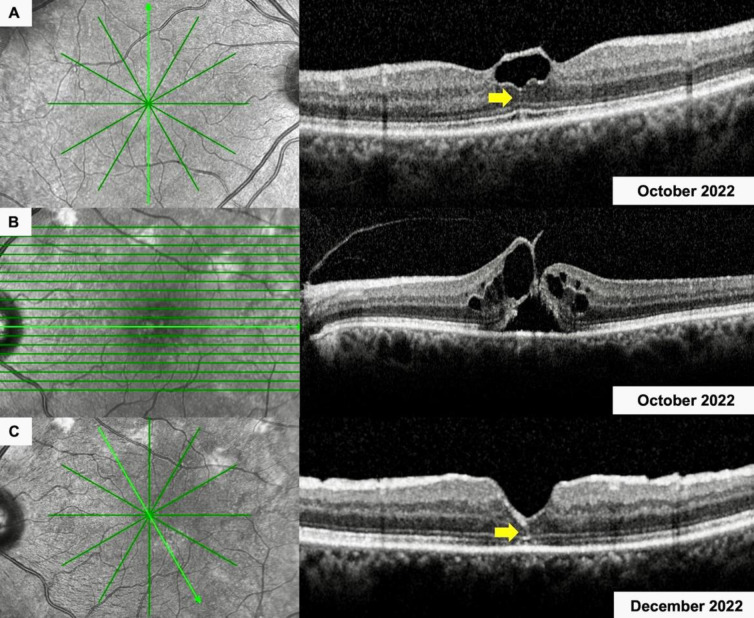
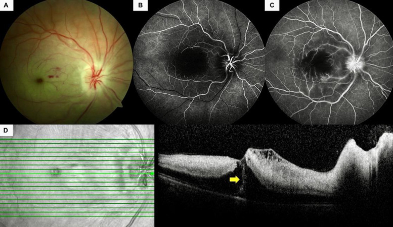
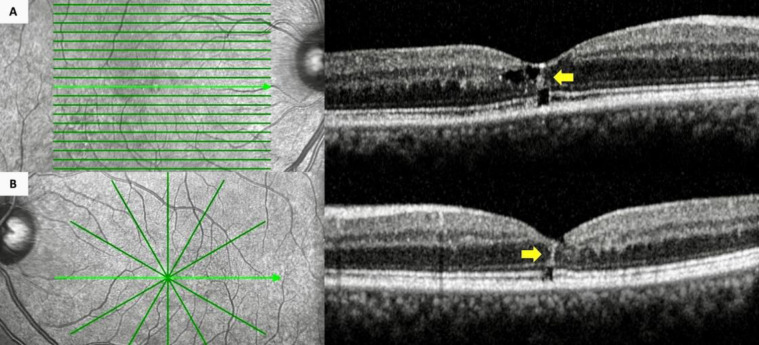
Background: The intraretinal hyperreflective line (IHL) is a novel posterior segment finding demonstrable using careful optical coherence tomography (OCT) examination. It likely indicates a reaction against photoreceptor, Muller cell, and/or retinal pigment epithelial damage. This study analyzed the spectral-domain OCT characteristics of IHLs to disclose their presence in various retinal conditions.
Methods: A retrospective review of the charted and imaging records of participants with IHL was conducted at Dokuz Eylul University Department of Ophthalmology between January 2019 and August 2023. The inclusion criterion was the detection of an IHL on good-quality B-scan spectral-domain OCT. An IHL was defined as a vertical line extending from the ellipsoid zone band (or lower) through the outer nuclear layer to the internal limiting membrane in the central fovea. Associated retinal conditions were recorded as potential causative factors for the presence of IHL.
Results: IHL was observed on spectral-domain OCT in 40 eyes of 38 participants with several retinal diseases assessment. Fourteen eyes (35%) underwent vitreoretinal surgery pre-IHL detection (12 were operated for full-thickness macular hole [FTMH], one for epiretinal membrane [ERM], and one for rhegmatogenous retinal detachment). In six eyes (15%) a microhole coexisted. Four eyes (10%) had a concurrent lamellar macular hole. The IHL preceded the occurrence of FTMH in three eyes (7.5%), and diabetic macular edema and type 2 idiopathic macular telangiectasia (MacTel-2) were present in three eyes (7.5%) each. The remaining conditions included vitreomacular traction (VMT), nonarteritic anterior ischemic optic neuropathy with central retinal artery occlusion, commotio retinae, exudative age-related macular degeneration, ERM, non-infectious idiopathic posterior uveitis, and Coats' disease, each affecting one eye (2.5%). Of the two participants with bilateral involvement, one was diagnosed with MacTel-2 and the other had IHL with VMT in the right eye that was detected post-vitreoretinal surgery for FTMH in the left eye.
Conclusions: Although IHLs are mostly identified in eyes with vitreomacular surface diseases, clinicians may encounter IHLs in other types of retinal pathology. Further large-scale, multicenter, long-term studies on the presence of IHLs in OCT imaging are required to provide more substantial insight on this biomarker.


 分享
分享
 求助内容:
求助内容: 应助结果提醒方式:
 扫码关注我们
