Mateo Gende , Joaquim de Moura , Patricia Robles , Jose Fernández-Vigo , José M. Martínez-de-la-Casa , GlaucoClub AI, Julián García-Feijóo , Jorge Novo , Marcos Ortega
{"title":"基于环状毛细血管 OCT 的青光眼和高度近视患者视网膜层厚度多扇区分析。","authors":"Mateo Gende , Joaquim de Moura , Patricia Robles , Jose Fernández-Vigo , José M. Martínez-de-la-Casa , GlaucoClub AI, Julián García-Feijóo , Jorge Novo , Marcos Ortega","doi":"10.1016/j.compmedimag.2024.102464","DOIUrl":null,"url":null,"abstract":"<div><div>Glaucoma is the leading cause of irreversible blindness worldwide. The diagnosis process for glaucoma involves the measurement of the thickness of retinal layers in order to track its degeneration. The elongated shape of highly myopic eyes can hinder this diagnosis process, since it affects the OCT scanning process, producing deformations that can mimic or mask the degeneration caused by glaucoma. In this work, we present the first comprehensive cross-disease analysis that is focused on the anatomical structures most impacted in glaucoma and high myopia patients, facilitating precise differential diagnosis from those solely afflicted by myopia. To achieve this, a fully automatic approach for the retinal layer segmentation was specifically tailored for the accurate measurement of retinal thickness in both highly myopic and emmetropic eyes. To the best of our knowledge, this is the first approach proposed for the analysis of retinal layers in circumpapillary optical coherence tomography images that takes into account the elongation of the eyes in myopia, thus addressing critical diagnostic needs. The results from this study indicate that the temporal superior (mean difference <span><math><mrow><mn>11</mn><mo>.</mo><mn>1</mn><mspace></mspace><mi>μ</mi><mi>m</mi></mrow></math></span>, <span><math><mrow><mi>p</mi><mo><</mo><mn>0</mn><mo>.</mo><mn>05</mn></mrow></math></span>), nasal inferior (<span><math><mrow><mn>13</mn><mo>.</mo><mn>1</mn><mspace></mspace><mi>μ</mi><mi>m</mi></mrow></math></span>, <span><math><mrow><mi>p</mi><mo><</mo><mn>0</mn><mo>.</mo><mn>01</mn></mrow></math></span>) and temporal inferior (<span><math><mrow><mn>13</mn><mo>.</mo><mn>3</mn><mspace></mspace><mi>μ</mi><mi>m</mi></mrow></math></span>, <span><math><mrow><mi>p</mi><mo><</mo><mn>0</mn><mo>.</mo><mn>01</mn></mrow></math></span>) sectors of the retinal nerve fibre layer show the most significant reduction in retinal thickness in patients of glaucoma and myopia with regards to patients of myopia.</div></div>","PeriodicalId":50631,"journal":{"name":"Computerized Medical Imaging and Graphics","volume":"118 ","pages":"Article 102464"},"PeriodicalIF":5.5000,"publicationDate":"2024-12-01","publicationTypes":"Journal Article","fieldsOfStudy":null,"isOpenAccess":false,"openAccessPdf":"","citationCount":"0","resultStr":"{\"title\":\"Circumpapillary OCT-based multi-sector analysis of retinal layer thickness in patients with glaucoma and high myopia\",\"authors\":\"Mateo Gende , Joaquim de Moura , Patricia Robles , Jose Fernández-Vigo , José M. Martínez-de-la-Casa , GlaucoClub AI, Julián García-Feijóo , Jorge Novo , Marcos Ortega\",\"doi\":\"10.1016/j.compmedimag.2024.102464\",\"DOIUrl\":null,\"url\":null,\"abstract\":\"<div><div>Glaucoma is the leading cause of irreversible blindness worldwide. The diagnosis process for glaucoma involves the measurement of the thickness of retinal layers in order to track its degeneration. The elongated shape of highly myopic eyes can hinder this diagnosis process, since it affects the OCT scanning process, producing deformations that can mimic or mask the degeneration caused by glaucoma. In this work, we present the first comprehensive cross-disease analysis that is focused on the anatomical structures most impacted in glaucoma and high myopia patients, facilitating precise differential diagnosis from those solely afflicted by myopia. To achieve this, a fully automatic approach for the retinal layer segmentation was specifically tailored for the accurate measurement of retinal thickness in both highly myopic and emmetropic eyes. To the best of our knowledge, this is the first approach proposed for the analysis of retinal layers in circumpapillary optical coherence tomography images that takes into account the elongation of the eyes in myopia, thus addressing critical diagnostic needs. The results from this study indicate that the temporal superior (mean difference <span><math><mrow><mn>11</mn><mo>.</mo><mn>1</mn><mspace></mspace><mi>μ</mi><mi>m</mi></mrow></math></span>, <span><math><mrow><mi>p</mi><mo><</mo><mn>0</mn><mo>.</mo><mn>05</mn></mrow></math></span>), nasal inferior (<span><math><mrow><mn>13</mn><mo>.</mo><mn>1</mn><mspace></mspace><mi>μ</mi><mi>m</mi></mrow></math></span>, <span><math><mrow><mi>p</mi><mo><</mo><mn>0</mn><mo>.</mo><mn>01</mn></mrow></math></span>) and temporal inferior (<span><math><mrow><mn>13</mn><mo>.</mo><mn>3</mn><mspace></mspace><mi>μ</mi><mi>m</mi></mrow></math></span>, <span><math><mrow><mi>p</mi><mo><</mo><mn>0</mn><mo>.</mo><mn>01</mn></mrow></math></span>) sectors of the retinal nerve fibre layer show the most significant reduction in retinal thickness in patients of glaucoma and myopia with regards to patients of myopia.</div></div>\",\"PeriodicalId\":50631,\"journal\":{\"name\":\"Computerized Medical Imaging and Graphics\",\"volume\":\"118 \",\"pages\":\"Article 102464\"},\"PeriodicalIF\":5.5000,\"publicationDate\":\"2024-12-01\",\"publicationTypes\":\"Journal Article\",\"fieldsOfStudy\":null,\"isOpenAccess\":false,\"openAccessPdf\":\"\",\"citationCount\":\"0\",\"resultStr\":null,\"platform\":\"Semanticscholar\",\"paperid\":null,\"PeriodicalName\":\"Computerized Medical Imaging and Graphics\",\"FirstCategoryId\":\"5\",\"ListUrlMain\":\"https://www.sciencedirect.com/science/article/pii/S0895611124001411\",\"RegionNum\":2,\"RegionCategory\":\"医学\",\"ArticlePicture\":[],\"TitleCN\":null,\"AbstractTextCN\":null,\"PMCID\":null,\"EPubDate\":\"2024/11/19 0:00:00\",\"PubModel\":\"Epub\",\"JCR\":\"Q1\",\"JCRName\":\"ENGINEERING, BIOMEDICAL\",\"Score\":null,\"Total\":0}","platform":"Semanticscholar","paperid":null,"PeriodicalName":"Computerized Medical Imaging and Graphics","FirstCategoryId":"5","ListUrlMain":"https://www.sciencedirect.com/science/article/pii/S0895611124001411","RegionNum":2,"RegionCategory":"医学","ArticlePicture":[],"TitleCN":null,"AbstractTextCN":null,"PMCID":null,"EPubDate":"2024/11/19 0:00:00","PubModel":"Epub","JCR":"Q1","JCRName":"ENGINEERING, BIOMEDICAL","Score":null,"Total":0}
引用次数: 0
摘要
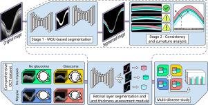
青光眼是导致全球不可逆失明的主要原因。青光眼的诊断过程包括测量视网膜层的厚度,以跟踪其退化情况。高度近视眼的细长形状会影响 OCT 扫描过程,产生变形,从而模仿或掩盖青光眼引起的变性,因此会阻碍诊断过程。在这项工作中,我们首次提出了全面的跨疾病分析,重点关注青光眼和高度近视患者中受影响最大的解剖结构,从而有助于与单纯近视患者进行精确的鉴别诊断。为此,我们专门定制了一种全自动视网膜层分割方法,用于精确测量高度近视眼和弱视眼的视网膜厚度。据我们所知,这是第一种用于分析环状毛细血管光学相干断层扫描图像中视网膜层的方法,它考虑到了近视眼的眼球拉长问题,从而满足了重要的诊断需求。这项研究的结果表明,颞上部(平均差 11.1μm,p
Circumpapillary OCT-based multi-sector analysis of retinal layer thickness in patients with glaucoma and high myopia
Glaucoma is the leading cause of irreversible blindness worldwide. The diagnosis process for glaucoma involves the measurement of the thickness of retinal layers in order to track its degeneration. The elongated shape of highly myopic eyes can hinder this diagnosis process, since it affects the OCT scanning process, producing deformations that can mimic or mask the degeneration caused by glaucoma. In this work, we present the first comprehensive cross-disease analysis that is focused on the anatomical structures most impacted in glaucoma and high myopia patients, facilitating precise differential diagnosis from those solely afflicted by myopia. To achieve this, a fully automatic approach for the retinal layer segmentation was specifically tailored for the accurate measurement of retinal thickness in both highly myopic and emmetropic eyes. To the best of our knowledge, this is the first approach proposed for the analysis of retinal layers in circumpapillary optical coherence tomography images that takes into account the elongation of the eyes in myopia, thus addressing critical diagnostic needs. The results from this study indicate that the temporal superior (mean difference , ), nasal inferior (, ) and temporal inferior (, ) sectors of the retinal nerve fibre layer show the most significant reduction in retinal thickness in patients of glaucoma and myopia with regards to patients of myopia.
期刊介绍:
The purpose of the journal Computerized Medical Imaging and Graphics is to act as a source for the exchange of research results concerning algorithmic advances, development, and application of digital imaging in disease detection, diagnosis, intervention, prevention, precision medicine, and population health. Included in the journal will be articles on novel computerized imaging or visualization techniques, including artificial intelligence and machine learning, augmented reality for surgical planning and guidance, big biomedical data visualization, computer-aided diagnosis, computerized-robotic surgery, image-guided therapy, imaging scanning and reconstruction, mobile and tele-imaging, radiomics, and imaging integration and modeling with other information relevant to digital health. The types of biomedical imaging include: magnetic resonance, computed tomography, ultrasound, nuclear medicine, X-ray, microwave, optical and multi-photon microscopy, video and sensory imaging, and the convergence of biomedical images with other non-imaging datasets.


 分享
分享
 求助内容:
求助内容: 应助结果提醒方式:
 扫码关注我们
