Zinhle P Mlambo, Motshedisi Sebitloane, Thajasvarie Naicker
{"title":"胎盘生长因子 (PlGF) 和可溶性 FMS 样酪氨酸激酶 1 (sFlt-1) 在感染 HIV 的非洲裔先兆子痫妇女的胎盘床中的免疫表达。","authors":"Zinhle P Mlambo, Motshedisi Sebitloane, Thajasvarie Naicker","doi":"10.1007/s00418-024-02341-6","DOIUrl":null,"url":null,"abstract":"<p><p>Preeclampsia, a severe pregnancy complication linked to defective placentation, poses significant maternal risks and is characterized by dysregulated angiogenic factors, including placental growth factor (PlGF) and soluble fms-like tyrosine kinase-1 (sFlt-1). Women with HIV/AIDS and receiving ART may face an increased susceptibility to preeclampsia development due to immunological and angiogenic imbalance. This study investigates the immunoexpression of these factors in the context of HIV-associated preeclampsia, utilizing morphometric image analysis. The study cohort comprised 180 women, including 60 normotensive and 120 preeclamptic participants, further stratified by HIV status and gestational age (early-onset PE [EOPE] < 34 weeks and late-onset PE [LOPE] ≥ 34 weeks). Placental bed tissues were immunostained with mouse anti-human sFlt-1 and PlGF antibodies, and the results were analyzed using Zeiss Axio-Vision and GraphPad Prism software. sFlt-1 levels showed no significant overall difference between preeclamptic and normotensive women (p = 0.8661), though slightly increased in the preeclamptic myometrium, independent of HIV status. However, sFlt-1 levels were significantly higher in EOPE compared to both normotensive and LOPE groups. PlGF immunostaining also showed no significant overall difference (p = 0.7387) but was notably lower in preeclamptic pregnancies and significantly higher in EOPE compared to LOPE. HIV status did not significantly impact sFlt-1 or PlGF levels, although sFlt-1 was slightly higher in HIV-negative women, while PlGF was marginally higher in HIV-positive women. These findings highlight the complex role of angiogenic factors in preeclampsia pathophysiology and suggest that antiretroviral therapies (ARTs) may contribute to the dysregulation of these factors due to a heightened immune milieu.</p>","PeriodicalId":13107,"journal":{"name":"Histochemistry and Cell Biology","volume":"163 1","pages":"8"},"PeriodicalIF":2.1000,"publicationDate":"2024-11-23","publicationTypes":"Journal Article","fieldsOfStudy":null,"isOpenAccess":false,"openAccessPdf":"https://www.ncbi.nlm.nih.gov/pmc/articles/PMC11585514/pdf/","citationCount":"0","resultStr":"{\"title\":\"Immunoexpression of placental growth factor (PlGF) and soluble FMS-like tyrosine kinase 1 (sFlt-1) in the placental bed of preeclamptic women of African ancestry living with HIV infection.\",\"authors\":\"Zinhle P Mlambo, Motshedisi Sebitloane, Thajasvarie Naicker\",\"doi\":\"10.1007/s00418-024-02341-6\",\"DOIUrl\":null,\"url\":null,\"abstract\":\"<p><p>Preeclampsia, a severe pregnancy complication linked to defective placentation, poses significant maternal risks and is characterized by dysregulated angiogenic factors, including placental growth factor (PlGF) and soluble fms-like tyrosine kinase-1 (sFlt-1). Women with HIV/AIDS and receiving ART may face an increased susceptibility to preeclampsia development due to immunological and angiogenic imbalance. This study investigates the immunoexpression of these factors in the context of HIV-associated preeclampsia, utilizing morphometric image analysis. The study cohort comprised 180 women, including 60 normotensive and 120 preeclamptic participants, further stratified by HIV status and gestational age (early-onset PE [EOPE] < 34 weeks and late-onset PE [LOPE] ≥ 34 weeks). Placental bed tissues were immunostained with mouse anti-human sFlt-1 and PlGF antibodies, and the results were analyzed using Zeiss Axio-Vision and GraphPad Prism software. sFlt-1 levels showed no significant overall difference between preeclamptic and normotensive women (p = 0.8661), though slightly increased in the preeclamptic myometrium, independent of HIV status. However, sFlt-1 levels were significantly higher in EOPE compared to both normotensive and LOPE groups. PlGF immunostaining also showed no significant overall difference (p = 0.7387) but was notably lower in preeclamptic pregnancies and significantly higher in EOPE compared to LOPE. HIV status did not significantly impact sFlt-1 or PlGF levels, although sFlt-1 was slightly higher in HIV-negative women, while PlGF was marginally higher in HIV-positive women. These findings highlight the complex role of angiogenic factors in preeclampsia pathophysiology and suggest that antiretroviral therapies (ARTs) may contribute to the dysregulation of these factors due to a heightened immune milieu.</p>\",\"PeriodicalId\":13107,\"journal\":{\"name\":\"Histochemistry and Cell Biology\",\"volume\":\"163 1\",\"pages\":\"8\"},\"PeriodicalIF\":2.1000,\"publicationDate\":\"2024-11-23\",\"publicationTypes\":\"Journal Article\",\"fieldsOfStudy\":null,\"isOpenAccess\":false,\"openAccessPdf\":\"https://www.ncbi.nlm.nih.gov/pmc/articles/PMC11585514/pdf/\",\"citationCount\":\"0\",\"resultStr\":null,\"platform\":\"Semanticscholar\",\"paperid\":null,\"PeriodicalName\":\"Histochemistry and Cell Biology\",\"FirstCategoryId\":\"99\",\"ListUrlMain\":\"https://doi.org/10.1007/s00418-024-02341-6\",\"RegionNum\":4,\"RegionCategory\":\"生物学\",\"ArticlePicture\":[],\"TitleCN\":null,\"AbstractTextCN\":null,\"PMCID\":null,\"EPubDate\":\"\",\"PubModel\":\"\",\"JCR\":\"Q4\",\"JCRName\":\"CELL BIOLOGY\",\"Score\":null,\"Total\":0}","platform":"Semanticscholar","paperid":null,"PeriodicalName":"Histochemistry and Cell Biology","FirstCategoryId":"99","ListUrlMain":"https://doi.org/10.1007/s00418-024-02341-6","RegionNum":4,"RegionCategory":"生物学","ArticlePicture":[],"TitleCN":null,"AbstractTextCN":null,"PMCID":null,"EPubDate":"","PubModel":"","JCR":"Q4","JCRName":"CELL BIOLOGY","Score":null,"Total":0}
引用次数: 0
摘要
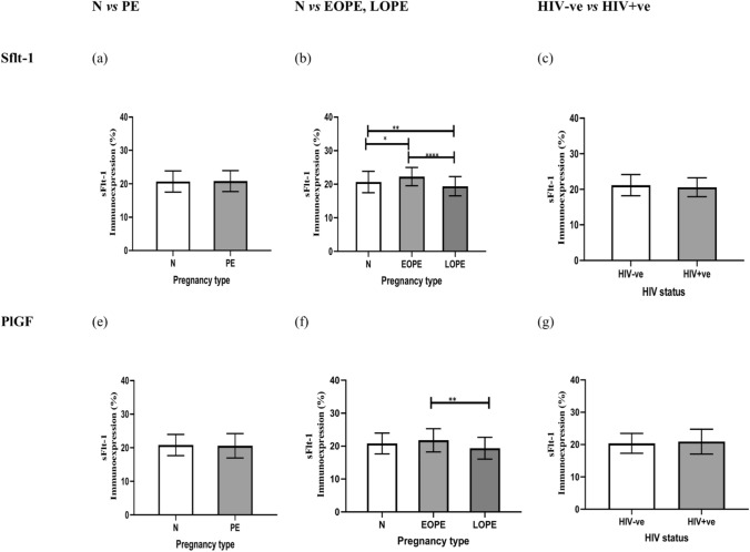
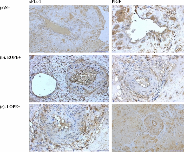
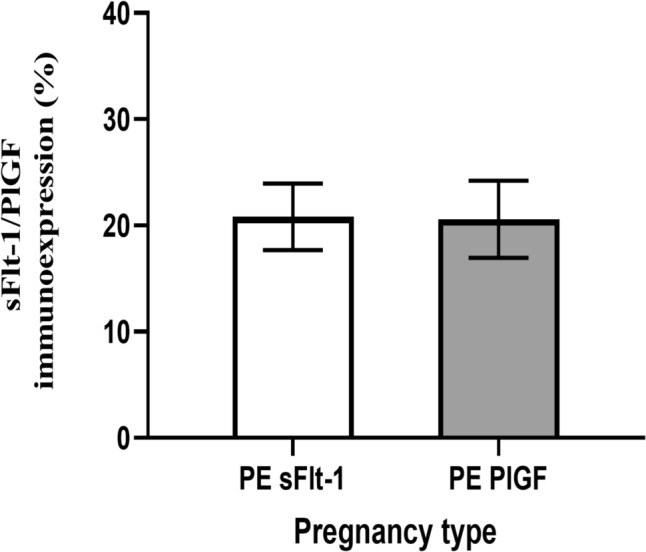
子痫前期是一种与胎盘功能缺陷有关的严重妊娠并发症,对孕产妇的风险很大,其特点是血管生成因子失调,包括胎盘生长因子(PlGF)和可溶性 fms 样酪氨酸激酶-1(sFlt-1)。感染艾滋病毒/艾滋病并接受抗逆转录病毒疗法的妇女可能会因免疫和血管生成失衡而更容易发生子痫前期。本研究利用形态计量学图像分析,调查了这些因子在 HIV 相关子痫前期中的免疫表达情况。研究队列由 180 名妇女组成,其中包括 60 名血压正常者和 120 名子痫前期患者,并根据 HIV 感染状况和胎龄进行了进一步分层(早发 PE [EOPE] 和晚发 PE [EOPE] )。
Immunoexpression of placental growth factor (PlGF) and soluble FMS-like tyrosine kinase 1 (sFlt-1) in the placental bed of preeclamptic women of African ancestry living with HIV infection.
Preeclampsia, a severe pregnancy complication linked to defective placentation, poses significant maternal risks and is characterized by dysregulated angiogenic factors, including placental growth factor (PlGF) and soluble fms-like tyrosine kinase-1 (sFlt-1). Women with HIV/AIDS and receiving ART may face an increased susceptibility to preeclampsia development due to immunological and angiogenic imbalance. This study investigates the immunoexpression of these factors in the context of HIV-associated preeclampsia, utilizing morphometric image analysis. The study cohort comprised 180 women, including 60 normotensive and 120 preeclamptic participants, further stratified by HIV status and gestational age (early-onset PE [EOPE] < 34 weeks and late-onset PE [LOPE] ≥ 34 weeks). Placental bed tissues were immunostained with mouse anti-human sFlt-1 and PlGF antibodies, and the results were analyzed using Zeiss Axio-Vision and GraphPad Prism software. sFlt-1 levels showed no significant overall difference between preeclamptic and normotensive women (p = 0.8661), though slightly increased in the preeclamptic myometrium, independent of HIV status. However, sFlt-1 levels were significantly higher in EOPE compared to both normotensive and LOPE groups. PlGF immunostaining also showed no significant overall difference (p = 0.7387) but was notably lower in preeclamptic pregnancies and significantly higher in EOPE compared to LOPE. HIV status did not significantly impact sFlt-1 or PlGF levels, although sFlt-1 was slightly higher in HIV-negative women, while PlGF was marginally higher in HIV-positive women. These findings highlight the complex role of angiogenic factors in preeclampsia pathophysiology and suggest that antiretroviral therapies (ARTs) may contribute to the dysregulation of these factors due to a heightened immune milieu.
期刊介绍:
Histochemistry and Cell Biology is devoted to the field of molecular histology and cell biology, publishing original articles dealing with the localization and identification of molecular components, metabolic activities and cell biological aspects of cells and tissues. Coverage extends to the development, application, and/or evaluation of methods and probes that can be used in the entire area of histochemistry and cell biology.


 分享
分享
 求助内容:
求助内容: 应助结果提醒方式:
 扫码关注我们
