Nan Yi, Lingyun Zhang, Xiangbo Huang, Jilei Ma, Jian Gao
{"title":"伦伐替尼激活的NDUFA4L2/IL33/PADI4通路可诱导中性粒细胞胞外陷阱,从而抑制肝细胞癌的杯突症。","authors":"Nan Yi, Lingyun Zhang, Xiangbo Huang, Jilei Ma, Jian Gao","doi":"10.1007/s13402-024-01013-w","DOIUrl":null,"url":null,"abstract":"<p><strong>Background: </strong>Lenvatinib is a potent first-line therapy for patients with hepatocellular carcinoma (HCC), but it also increased the number of neutrophils in HCC tumor microenvironment.</p><p><strong>Methods: </strong>CitH3, MPO-DNA, elastase and MPO activity were measured for assessing neutrophil extracellular traps (NETs) in vivo and in vitro. Cell cuproptosis was assessed by measurement of copper content, FDX1, and pyruvate. The functions of lenvatinib, DNase I, interleukin 33 (IL33) neutralizing antibody and GPX4 in tumor growth were explored in mice.</p><p><strong>Results: </strong>Lenvatinib induced NETs in the HCC tumor microenvironment via HCC cells, but not through the direct stimulation of neutrophils. In addition, NET clearance by DNase I improves the efficacy of lenvatinib therapy in HCC mouse models. Mechanistically, lenvatinib promoted the expression and secretion of IL33 by HCC cells that triggered NET formation. Moreover, IL33 knockdown in Hepa1-6 cells improved lenvatinib efficacy in Hepa1-6-bearing HCC model mice and reduced NET formation in the tumor microenvironment. Subsequently, lenvatinib increased IL33 production by increasing the NDUFA4L2 expression in HCC cells. Furthermore, we found that IL33 triggered NET formation in neutrophils by increasing the protein expression of PADI4 via the Akt/mTOR signaling pathway. Rapamycin inhibition of mTOR reduced PADI4 expression and NET formation. Consistently, PADI4 inhibition by the selective PAD4 inhibitor GSK484 hydrochloride (GSK484) improved lenvatinib response to HCC therapy. Importantly, NETs contribute to lenvatinib resistance by inhibiting cuproptosis, but not apoptosis, pyroptosis, or ferroptosis in HCC cells. Treatment with GSK484 reversed the inhibitory effects of NETs on cuproptosis and sensitized the HCC cells to lenvatinib.</p><p><strong>Conclusions: </strong>Our study revealed that lenvatinib-induced NETs inhibited the cuproptosis of HCC cells, suggesting that targeting the IL33/PADI4/NET axis represents a promising therapeutic strategy for ameliorating lenvatinib resistance in HCC.</p>","PeriodicalId":49223,"journal":{"name":"Cellular Oncology","volume":" ","pages":"487-504"},"PeriodicalIF":4.8000,"publicationDate":"2025-04-01","publicationTypes":"Journal Article","fieldsOfStudy":null,"isOpenAccess":false,"openAccessPdf":"https://www.ncbi.nlm.nih.gov/pmc/articles/PMC11996955/pdf/","citationCount":"0","resultStr":"{\"title\":\"Lenvatinib-activated NDUFA4L2/IL33/PADI4 pathway induces neutrophil extracellular traps that inhibit cuproptosis in hepatocellular carcinoma.\",\"authors\":\"Nan Yi, Lingyun Zhang, Xiangbo Huang, Jilei Ma, Jian Gao\",\"doi\":\"10.1007/s13402-024-01013-w\",\"DOIUrl\":null,\"url\":null,\"abstract\":\"<p><strong>Background: </strong>Lenvatinib is a potent first-line therapy for patients with hepatocellular carcinoma (HCC), but it also increased the number of neutrophils in HCC tumor microenvironment.</p><p><strong>Methods: </strong>CitH3, MPO-DNA, elastase and MPO activity were measured for assessing neutrophil extracellular traps (NETs) in vivo and in vitro. Cell cuproptosis was assessed by measurement of copper content, FDX1, and pyruvate. The functions of lenvatinib, DNase I, interleukin 33 (IL33) neutralizing antibody and GPX4 in tumor growth were explored in mice.</p><p><strong>Results: </strong>Lenvatinib induced NETs in the HCC tumor microenvironment via HCC cells, but not through the direct stimulation of neutrophils. In addition, NET clearance by DNase I improves the efficacy of lenvatinib therapy in HCC mouse models. Mechanistically, lenvatinib promoted the expression and secretion of IL33 by HCC cells that triggered NET formation. Moreover, IL33 knockdown in Hepa1-6 cells improved lenvatinib efficacy in Hepa1-6-bearing HCC model mice and reduced NET formation in the tumor microenvironment. Subsequently, lenvatinib increased IL33 production by increasing the NDUFA4L2 expression in HCC cells. Furthermore, we found that IL33 triggered NET formation in neutrophils by increasing the protein expression of PADI4 via the Akt/mTOR signaling pathway. Rapamycin inhibition of mTOR reduced PADI4 expression and NET formation. Consistently, PADI4 inhibition by the selective PAD4 inhibitor GSK484 hydrochloride (GSK484) improved lenvatinib response to HCC therapy. Importantly, NETs contribute to lenvatinib resistance by inhibiting cuproptosis, but not apoptosis, pyroptosis, or ferroptosis in HCC cells. Treatment with GSK484 reversed the inhibitory effects of NETs on cuproptosis and sensitized the HCC cells to lenvatinib.</p><p><strong>Conclusions: </strong>Our study revealed that lenvatinib-induced NETs inhibited the cuproptosis of HCC cells, suggesting that targeting the IL33/PADI4/NET axis represents a promising therapeutic strategy for ameliorating lenvatinib resistance in HCC.</p>\",\"PeriodicalId\":49223,\"journal\":{\"name\":\"Cellular Oncology\",\"volume\":\" \",\"pages\":\"487-504\"},\"PeriodicalIF\":4.8000,\"publicationDate\":\"2025-04-01\",\"publicationTypes\":\"Journal Article\",\"fieldsOfStudy\":null,\"isOpenAccess\":false,\"openAccessPdf\":\"https://www.ncbi.nlm.nih.gov/pmc/articles/PMC11996955/pdf/\",\"citationCount\":\"0\",\"resultStr\":null,\"platform\":\"Semanticscholar\",\"paperid\":null,\"PeriodicalName\":\"Cellular Oncology\",\"FirstCategoryId\":\"3\",\"ListUrlMain\":\"https://doi.org/10.1007/s13402-024-01013-w\",\"RegionNum\":2,\"RegionCategory\":\"医学\",\"ArticlePicture\":[],\"TitleCN\":null,\"AbstractTextCN\":null,\"PMCID\":null,\"EPubDate\":\"2024/11/25 0:00:00\",\"PubModel\":\"Epub\",\"JCR\":\"Q2\",\"JCRName\":\"CELL BIOLOGY\",\"Score\":null,\"Total\":0}","platform":"Semanticscholar","paperid":null,"PeriodicalName":"Cellular Oncology","FirstCategoryId":"3","ListUrlMain":"https://doi.org/10.1007/s13402-024-01013-w","RegionNum":2,"RegionCategory":"医学","ArticlePicture":[],"TitleCN":null,"AbstractTextCN":null,"PMCID":null,"EPubDate":"2024/11/25 0:00:00","PubModel":"Epub","JCR":"Q2","JCRName":"CELL BIOLOGY","Score":null,"Total":0}
引用次数: 0
摘要
背景伦伐替尼是肝细胞癌(HCC)患者的有效一线疗法,但它也会增加HCC肿瘤微环境中的中性粒细胞数量:方法:测量CitH3、MPO-DNA、弹性蛋白酶和MPO活性,以评估体内和体外的中性粒细胞胞外陷阱(NET)。通过测量铜含量、FDX1和丙酮酸来评估细胞杯突。在小鼠体内探讨了来伐替尼、DNase I、白细胞介素33(IL33)中和抗体和GPX4在肿瘤生长中的功能:结果:来伐替尼通过HCC细胞诱导HCC肿瘤微环境中的NET,而不是通过直接刺激中性粒细胞。此外,通过DNase I清除NET可提高来伐替尼治疗HCC小鼠模型的疗效。从机制上讲,来伐替尼促进了HCC细胞表达和分泌IL33,从而引发NET的形成。此外,敲除Hepa1-6细胞中的IL33可提高来伐替尼在Hepa1-6携带的HCC模型小鼠中的疗效,并减少肿瘤微环境中NET的形成。随后,来伐替尼通过增加HCC细胞中NDUFA4L2的表达来增加IL33的产生。此外,我们还发现IL33通过Akt/mTOR信号通路增加了PADI4的蛋白表达,从而引发了中性粒细胞中NET的形成。雷帕霉素抑制 mTOR 可减少 PADI4 的表达和 NET 的形成。同样,通过选择性PAD4抑制剂盐酸GSK484(GSK484)抑制PADI4可改善来伐替尼对HCC治疗的反应。重要的是,NETs通过抑制HCC细胞的杯突变而非凋亡、热突变或铁突变,从而导致来伐替尼耐药。用GSK484治疗可逆转NETs对杯突症的抑制作用,并使HCC细胞对来伐替尼敏感:我们的研究发现,来伐替尼诱导的NET抑制了HCC细胞的杯突症,这表明靶向IL33/PADI4/NET轴是改善HCC对来伐替尼耐药的一种很有前景的治疗策略。
Lenvatinib-activated NDUFA4L2/IL33/PADI4 pathway induces neutrophil extracellular traps that inhibit cuproptosis in hepatocellular carcinoma.
Background: Lenvatinib is a potent first-line therapy for patients with hepatocellular carcinoma (HCC), but it also increased the number of neutrophils in HCC tumor microenvironment.
Methods: CitH3, MPO-DNA, elastase and MPO activity were measured for assessing neutrophil extracellular traps (NETs) in vivo and in vitro. Cell cuproptosis was assessed by measurement of copper content, FDX1, and pyruvate. The functions of lenvatinib, DNase I, interleukin 33 (IL33) neutralizing antibody and GPX4 in tumor growth were explored in mice.
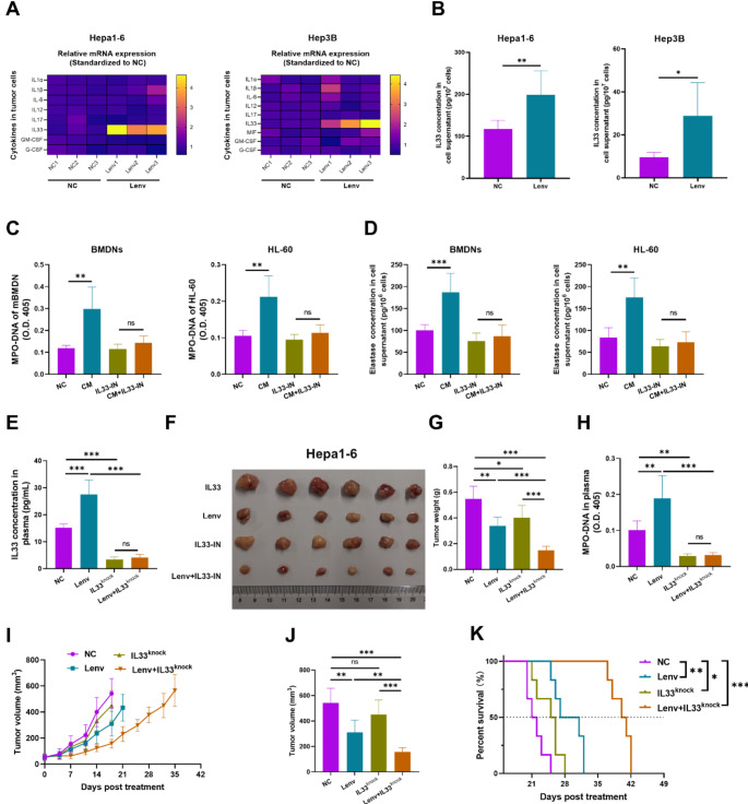
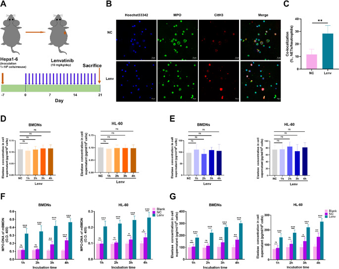
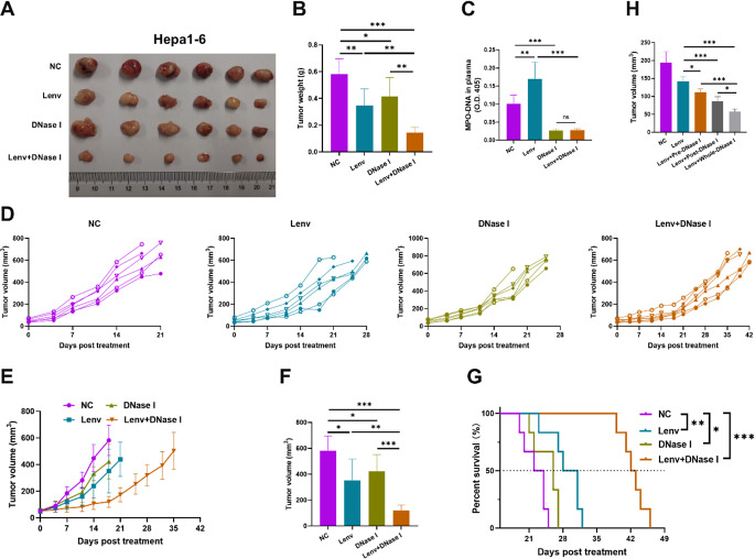
Results: Lenvatinib induced NETs in the HCC tumor microenvironment via HCC cells, but not through the direct stimulation of neutrophils. In addition, NET clearance by DNase I improves the efficacy of lenvatinib therapy in HCC mouse models. Mechanistically, lenvatinib promoted the expression and secretion of IL33 by HCC cells that triggered NET formation. Moreover, IL33 knockdown in Hepa1-6 cells improved lenvatinib efficacy in Hepa1-6-bearing HCC model mice and reduced NET formation in the tumor microenvironment. Subsequently, lenvatinib increased IL33 production by increasing the NDUFA4L2 expression in HCC cells. Furthermore, we found that IL33 triggered NET formation in neutrophils by increasing the protein expression of PADI4 via the Akt/mTOR signaling pathway. Rapamycin inhibition of mTOR reduced PADI4 expression and NET formation. Consistently, PADI4 inhibition by the selective PAD4 inhibitor GSK484 hydrochloride (GSK484) improved lenvatinib response to HCC therapy. Importantly, NETs contribute to lenvatinib resistance by inhibiting cuproptosis, but not apoptosis, pyroptosis, or ferroptosis in HCC cells. Treatment with GSK484 reversed the inhibitory effects of NETs on cuproptosis and sensitized the HCC cells to lenvatinib.
Conclusions: Our study revealed that lenvatinib-induced NETs inhibited the cuproptosis of HCC cells, suggesting that targeting the IL33/PADI4/NET axis represents a promising therapeutic strategy for ameliorating lenvatinib resistance in HCC.
期刊介绍:
The Official Journal of the International Society for Cellular Oncology
Focuses on translational research
Addresses the conversion of cell biology to clinical applications
Cellular Oncology publishes scientific contributions from various biomedical and clinical disciplines involved in basic and translational cancer research on the cell and tissue level, technical and bioinformatics developments in this area, and clinical applications. This includes a variety of fields like genome technology, micro-arrays and other high-throughput techniques, genomic instability, SNP, DNA methylation, signaling pathways, DNA organization, (sub)microscopic imaging, proteomics, bioinformatics, functional effects of genomics, drug design and development, molecular diagnostics and targeted cancer therapies, genotype-phenotype interactions.
A major goal is to translate the latest developments in these fields from the research laboratory into routine patient management. To this end Cellular Oncology forms a platform of scientific information exchange between molecular biologists and geneticists, technical developers, pathologists, (medical) oncologists and other clinicians involved in the management of cancer patients.
In vitro studies are preferentially supported by validations in tumor tissue with clinicopathological associations.


 分享
分享
 求助内容:
求助内容: 应助结果提醒方式:
 扫码关注我们
