Ayça Küpeli Çınar, Riza Serttas, Abdulkadir Can Çınar, Hande Güçlü, Suat Erdogan
{"title":"如图所示,橙皮甙能抑制 TGF-β2 诱导的视网膜色素上皮细胞增殖和上皮-间质转化","authors":"Ayça Küpeli Çınar, Riza Serttas, Abdulkadir Can Çınar, Hande Güçlü, Suat Erdogan","doi":"10.1007/s10735-024-10275-5","DOIUrl":null,"url":null,"abstract":"<div><p>This study investigates the potential therapeutic effects and molecular mechanisms of hesperidin treatment on cell migration and epithelial-mesenchymal transition, key stages of proliferative vitreoretinopathy (PVR). Human retinal pigment epithelial cells (ARPE-19) were treated with 10 ng/ml transforming growth factor-beta 2 (TGF-β2) alone or in combination with 1.56 μM hesperidin for 48 h. The impact of treatment on cell migration was evaluated using a wound healing assay. Apoptosis was assessed using DNA staining. mRNA and protein expression were evaluated using real-time PCR and Western blot, respectively. Hesperidin inhibits the proliferation and transformation of the cells by inducing apoptosis and reverses the cell morphology modified by TGF-β2. Hesperidin inhibits cell migration induced by TGF-β2. Upon treatment with hesperidin, the levels of mesenchymal markers upregulated by TGF-β2, such as MMP-1, -2, -9, fibronectin, α-SMA and the transcription factors Snail, Slug and ZEB-1, were downregulated. Conversely, the epithelial marker E-cadherin is upregulated with hesperidin treatment. Additionally, TIMP-1 and TIMP-2 expression levels, which are downregulated, increase with the treatment. These results suggest that hesperidin may inhibit the migration and EMT processes of RPE cells involved in the development of PVR, indicating its potential as a therapeutic agent for treating PVR.</p></div>","PeriodicalId":650,"journal":{"name":"Journal of Molecular Histology","volume":"56 1","pages":""},"PeriodicalIF":2.2000,"publicationDate":"2024-11-29","publicationTypes":"Journal Article","fieldsOfStudy":null,"isOpenAccess":false,"openAccessPdf":"","citationCount":"0","resultStr":"{\"title\":\"As shown hesperidin suppresses TGF-β2-induced proliferation and epithelial-mesenchymal transition of retinal pigment epithelial cells\",\"authors\":\"Ayça Küpeli Çınar, Riza Serttas, Abdulkadir Can Çınar, Hande Güçlü, Suat Erdogan\",\"doi\":\"10.1007/s10735-024-10275-5\",\"DOIUrl\":null,\"url\":null,\"abstract\":\"<div><p>This study investigates the potential therapeutic effects and molecular mechanisms of hesperidin treatment on cell migration and epithelial-mesenchymal transition, key stages of proliferative vitreoretinopathy (PVR). Human retinal pigment epithelial cells (ARPE-19) were treated with 10 ng/ml transforming growth factor-beta 2 (TGF-β2) alone or in combination with 1.56 μM hesperidin for 48 h. The impact of treatment on cell migration was evaluated using a wound healing assay. Apoptosis was assessed using DNA staining. mRNA and protein expression were evaluated using real-time PCR and Western blot, respectively. Hesperidin inhibits the proliferation and transformation of the cells by inducing apoptosis and reverses the cell morphology modified by TGF-β2. Hesperidin inhibits cell migration induced by TGF-β2. Upon treatment with hesperidin, the levels of mesenchymal markers upregulated by TGF-β2, such as MMP-1, -2, -9, fibronectin, α-SMA and the transcription factors Snail, Slug and ZEB-1, were downregulated. Conversely, the epithelial marker E-cadherin is upregulated with hesperidin treatment. Additionally, TIMP-1 and TIMP-2 expression levels, which are downregulated, increase with the treatment. These results suggest that hesperidin may inhibit the migration and EMT processes of RPE cells involved in the development of PVR, indicating its potential as a therapeutic agent for treating PVR.</p></div>\",\"PeriodicalId\":650,\"journal\":{\"name\":\"Journal of Molecular Histology\",\"volume\":\"56 1\",\"pages\":\"\"},\"PeriodicalIF\":2.2000,\"publicationDate\":\"2024-11-29\",\"publicationTypes\":\"Journal Article\",\"fieldsOfStudy\":null,\"isOpenAccess\":false,\"openAccessPdf\":\"\",\"citationCount\":\"0\",\"resultStr\":null,\"platform\":\"Semanticscholar\",\"paperid\":null,\"PeriodicalName\":\"Journal of Molecular Histology\",\"FirstCategoryId\":\"99\",\"ListUrlMain\":\"https://link.springer.com/article/10.1007/s10735-024-10275-5\",\"RegionNum\":4,\"RegionCategory\":\"生物学\",\"ArticlePicture\":[],\"TitleCN\":null,\"AbstractTextCN\":null,\"PMCID\":null,\"EPubDate\":\"\",\"PubModel\":\"\",\"JCR\":\"Q3\",\"JCRName\":\"CELL BIOLOGY\",\"Score\":null,\"Total\":0}","platform":"Semanticscholar","paperid":null,"PeriodicalName":"Journal of Molecular Histology","FirstCategoryId":"99","ListUrlMain":"https://link.springer.com/article/10.1007/s10735-024-10275-5","RegionNum":4,"RegionCategory":"生物学","ArticlePicture":[],"TitleCN":null,"AbstractTextCN":null,"PMCID":null,"EPubDate":"","PubModel":"","JCR":"Q3","JCRName":"CELL BIOLOGY","Score":null,"Total":0}
As shown hesperidin suppresses TGF-β2-induced proliferation and epithelial-mesenchymal transition of retinal pigment epithelial cells
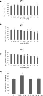
This study investigates the potential therapeutic effects and molecular mechanisms of hesperidin treatment on cell migration and epithelial-mesenchymal transition, key stages of proliferative vitreoretinopathy (PVR). Human retinal pigment epithelial cells (ARPE-19) were treated with 10 ng/ml transforming growth factor-beta 2 (TGF-β2) alone or in combination with 1.56 μM hesperidin for 48 h. The impact of treatment on cell migration was evaluated using a wound healing assay. Apoptosis was assessed using DNA staining. mRNA and protein expression were evaluated using real-time PCR and Western blot, respectively. Hesperidin inhibits the proliferation and transformation of the cells by inducing apoptosis and reverses the cell morphology modified by TGF-β2. Hesperidin inhibits cell migration induced by TGF-β2. Upon treatment with hesperidin, the levels of mesenchymal markers upregulated by TGF-β2, such as MMP-1, -2, -9, fibronectin, α-SMA and the transcription factors Snail, Slug and ZEB-1, were downregulated. Conversely, the epithelial marker E-cadherin is upregulated with hesperidin treatment. Additionally, TIMP-1 and TIMP-2 expression levels, which are downregulated, increase with the treatment. These results suggest that hesperidin may inhibit the migration and EMT processes of RPE cells involved in the development of PVR, indicating its potential as a therapeutic agent for treating PVR.
期刊介绍:
The Journal of Molecular Histology publishes results of original research on the localization and expression of molecules in animal cells, tissues and organs. Coverage includes studies describing novel cellular or ultrastructural distributions of molecules which provide insight into biochemical or physiological function, development, histologic structure and disease processes.
Major research themes of particular interest include:
- Cell-Cell and Cell-Matrix Interactions;
- Connective Tissues;
- Development and Disease;
- Neuroscience.
Please note that the Journal of Molecular Histology does not consider manuscripts dealing with the application of immunological or other probes on non-standard laboratory animal models unless the results are clearly of significant and general biological importance.
The Journal of Molecular Histology publishes full-length original research papers, review articles, short communications and letters to the editors. All manuscripts are typically reviewed by two independent referees. The Journal of Molecular Histology is a continuation of The Histochemical Journal.


 分享
分享
 求助内容:
求助内容: 应助结果提醒方式:
 扫码关注我们
