{"title":"口内吻合面部动静脉的解剖标志:尸体研究","authors":"Kengo Nakatsuka, Tomoyuki Yano, Takuya Omotehara, Shinichi Kawata, Masahiro Itoh","doi":"10.1002/micr.70004","DOIUrl":null,"url":null,"abstract":"<div>\n \n \n <section>\n \n <h3> Background</h3>\n \n <p>Intraoral anastomosis is a widely used technique for microvascular alveolar ridge augmentation and midface reconstruction. However, the predictable anatomical positioning of facial structures, such as the vessels, parotid duct, and facial nerve in the buccal region, has remained unclear. Therefore, we aimed to obtain the anatomical characteristics of these locations to establish surgical landmarks for the intraoral anastomosis of facial vessels.</p>\n </section>\n \n <section>\n \n <h3> Methods</h3>\n \n <p>A total of 26 sides from 13 formaldehyde-fixed cadavers approximately a month after fixation with a mean age at death of 86.6 ± 11.2 years (range: 55–104 years) were anatomically examined. Facial vessels, nerves, and the parotid duct were dissected intraorally. From the oral cavity side, the X-axis was defined as the line from the labial commissure to the lowest point of the intertragic notch.</p>\n </section>\n \n <section>\n \n <h3> Results</h3>\n \n <p>From the oral cavity side, all branches of the facial nerve were found under the facial artery and vein. The positioning order along the X-axis was the facial artery, vein, and parotid duct exit. The facial artery was 21.3 ± 2.2 mm and the facial vein was 39.2 ± 2.7 mm from the labial commissure. Ninety-two percent of facial veins were found within 15–20 mm of the facial artery on the X-axis. The parotid duct exit was 46.8 ± 2.0 mm from the labial commissure. In the buccal region, the vessel calibers of the facial artery and vein were 1.8 ± 0.2 and 2.1 ± 0.2 mm, respectively.</p>\n </section>\n \n <section>\n \n <h3> Conclusion</h3>\n \n <p>Knowledge of the anatomical relations among the facial artery, vein, parotid duct, and facial nerve from the oral cavity side can enhance the safety and efficacy of midface reconstruction surgeries involving intraoral anastomosis procedures.</p>\n </section>\n </div>","PeriodicalId":18600,"journal":{"name":"Microsurgery","volume":"45 1","pages":""},"PeriodicalIF":1.7000,"publicationDate":"2024-11-29","publicationTypes":"Journal Article","fieldsOfStudy":null,"isOpenAccess":false,"openAccessPdf":"https://onlinelibrary.wiley.com/doi/epdf/10.1002/micr.70004","citationCount":"0","resultStr":"{\"title\":\"Anatomical Landmarks of the Facial Artery and Vein for Intraoral Anastomosis: A Cadaveric Study\",\"authors\":\"Kengo Nakatsuka, Tomoyuki Yano, Takuya Omotehara, Shinichi Kawata, Masahiro Itoh\",\"doi\":\"10.1002/micr.70004\",\"DOIUrl\":null,\"url\":null,\"abstract\":\"<div>\\n \\n \\n <section>\\n \\n <h3> Background</h3>\\n \\n <p>Intraoral anastomosis is a widely used technique for microvascular alveolar ridge augmentation and midface reconstruction. However, the predictable anatomical positioning of facial structures, such as the vessels, parotid duct, and facial nerve in the buccal region, has remained unclear. Therefore, we aimed to obtain the anatomical characteristics of these locations to establish surgical landmarks for the intraoral anastomosis of facial vessels.</p>\\n </section>\\n \\n <section>\\n \\n <h3> Methods</h3>\\n \\n <p>A total of 26 sides from 13 formaldehyde-fixed cadavers approximately a month after fixation with a mean age at death of 86.6 ± 11.2 years (range: 55–104 years) were anatomically examined. Facial vessels, nerves, and the parotid duct were dissected intraorally. From the oral cavity side, the X-axis was defined as the line from the labial commissure to the lowest point of the intertragic notch.</p>\\n </section>\\n \\n <section>\\n \\n <h3> Results</h3>\\n \\n <p>From the oral cavity side, all branches of the facial nerve were found under the facial artery and vein. The positioning order along the X-axis was the facial artery, vein, and parotid duct exit. The facial artery was 21.3 ± 2.2 mm and the facial vein was 39.2 ± 2.7 mm from the labial commissure. Ninety-two percent of facial veins were found within 15–20 mm of the facial artery on the X-axis. The parotid duct exit was 46.8 ± 2.0 mm from the labial commissure. In the buccal region, the vessel calibers of the facial artery and vein were 1.8 ± 0.2 and 2.1 ± 0.2 mm, respectively.</p>\\n </section>\\n \\n <section>\\n \\n <h3> Conclusion</h3>\\n \\n <p>Knowledge of the anatomical relations among the facial artery, vein, parotid duct, and facial nerve from the oral cavity side can enhance the safety and efficacy of midface reconstruction surgeries involving intraoral anastomosis procedures.</p>\\n </section>\\n </div>\",\"PeriodicalId\":18600,\"journal\":{\"name\":\"Microsurgery\",\"volume\":\"45 1\",\"pages\":\"\"},\"PeriodicalIF\":1.7000,\"publicationDate\":\"2024-11-29\",\"publicationTypes\":\"Journal Article\",\"fieldsOfStudy\":null,\"isOpenAccess\":false,\"openAccessPdf\":\"https://onlinelibrary.wiley.com/doi/epdf/10.1002/micr.70004\",\"citationCount\":\"0\",\"resultStr\":null,\"platform\":\"Semanticscholar\",\"paperid\":null,\"PeriodicalName\":\"Microsurgery\",\"FirstCategoryId\":\"3\",\"ListUrlMain\":\"https://onlinelibrary.wiley.com/doi/10.1002/micr.70004\",\"RegionNum\":3,\"RegionCategory\":\"医学\",\"ArticlePicture\":[],\"TitleCN\":null,\"AbstractTextCN\":null,\"PMCID\":null,\"EPubDate\":\"\",\"PubModel\":\"\",\"JCR\":\"Q3\",\"JCRName\":\"SURGERY\",\"Score\":null,\"Total\":0}","platform":"Semanticscholar","paperid":null,"PeriodicalName":"Microsurgery","FirstCategoryId":"3","ListUrlMain":"https://onlinelibrary.wiley.com/doi/10.1002/micr.70004","RegionNum":3,"RegionCategory":"医学","ArticlePicture":[],"TitleCN":null,"AbstractTextCN":null,"PMCID":null,"EPubDate":"","PubModel":"","JCR":"Q3","JCRName":"SURGERY","Score":null,"Total":0}
Anatomical Landmarks of the Facial Artery and Vein for Intraoral Anastomosis: A Cadaveric Study
Background
Intraoral anastomosis is a widely used technique for microvascular alveolar ridge augmentation and midface reconstruction. However, the predictable anatomical positioning of facial structures, such as the vessels, parotid duct, and facial nerve in the buccal region, has remained unclear. Therefore, we aimed to obtain the anatomical characteristics of these locations to establish surgical landmarks for the intraoral anastomosis of facial vessels.
Methods
A total of 26 sides from 13 formaldehyde-fixed cadavers approximately a month after fixation with a mean age at death of 86.6 ± 11.2 years (range: 55–104 years) were anatomically examined. Facial vessels, nerves, and the parotid duct were dissected intraorally. From the oral cavity side, the X-axis was defined as the line from the labial commissure to the lowest point of the intertragic notch.
Results
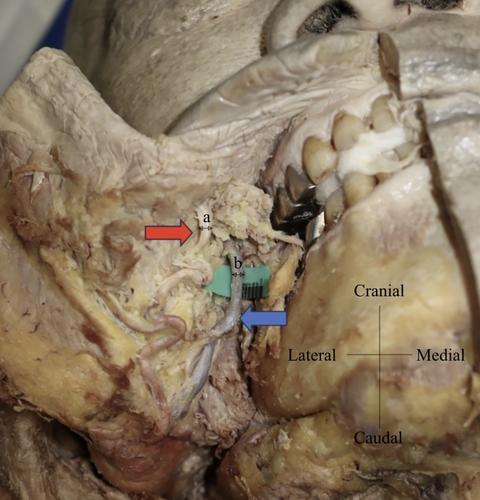
From the oral cavity side, all branches of the facial nerve were found under the facial artery and vein. The positioning order along the X-axis was the facial artery, vein, and parotid duct exit. The facial artery was 21.3 ± 2.2 mm and the facial vein was 39.2 ± 2.7 mm from the labial commissure. Ninety-two percent of facial veins were found within 15–20 mm of the facial artery on the X-axis. The parotid duct exit was 46.8 ± 2.0 mm from the labial commissure. In the buccal region, the vessel calibers of the facial artery and vein were 1.8 ± 0.2 and 2.1 ± 0.2 mm, respectively.
Conclusion
Knowledge of the anatomical relations among the facial artery, vein, parotid duct, and facial nerve from the oral cavity side can enhance the safety and efficacy of midface reconstruction surgeries involving intraoral anastomosis procedures.
期刊介绍:
Microsurgery is an international and interdisciplinary publication of original contributions concerning surgery under microscopic magnification. Microsurgery publishes clinical studies, research papers, invited articles, relevant reviews, and other scholarly works from all related fields including orthopaedic surgery, otolaryngology, pediatric surgery, plastic surgery, urology, and vascular surgery.


 分享
分享
 求助内容:
求助内容: 应助结果提醒方式:
 扫码关注我们
