Alexander Jans, Pieter Sinonquel, Tom C J Seerden, Alexander De Bodelier, Rogier de Ridder, Marieke J Pierik, John Gásdal Karstensen, Stine Sloth, Gert De Hertogh, Ingrid Demedts, Hilde Willekens, Severine Vermeire, Raf Bisschops
{"title":"染料染色内窥镜与i-scan虚拟染色内窥镜在长期溃疡性结肠炎中的比较:多中心前瞻性RCT。","authors":"Alexander Jans, Pieter Sinonquel, Tom C J Seerden, Alexander De Bodelier, Rogier de Ridder, Marieke J Pierik, John Gásdal Karstensen, Stine Sloth, Gert De Hertogh, Ingrid Demedts, Hilde Willekens, Severine Vermeire, Raf Bisschops","doi":"10.1055/a-2443-1080","DOIUrl":null,"url":null,"abstract":"<p><p><b>Background and study aims</b> Long-standing ulcerative colitis (UC) is associated with an increased risk of developing colorectal neoplasia. Both dye-based chromoendoscopy (DCE) and virtual chromoendoscopy (VCE) increase detection of neoplastic lesions. In this prospective randomized controlled trial (RCT), we compared the neoplasia detection rate between DCE and i-scan VCE in patients with long-standing UC. <b>Patient and methods</b> In four European hospitals, 131 patients with long-standing UC (disease duration > 8 years) were randomized to either DCE with methylene blue 0.1% (n = 66) or i-scan VCE (n = 65). All procedures were performed by trained endoscopists. Biopsies were taken from all visible lesions and the surrounding mucosa. <b>Results</b> The mean number of neoplastic lesions detected per colonoscopy was not significantly different between DCE (0.27) and i-scan VCE (0.37) ( <i>P</i> = 0.41). Similarly, there was no significant difference in neoplasia detection rate between DCE (19.7%) and VCE (27.7%) (odds ratio0.64, 95% confidence interval 0.28-1.50, <i>P</i> = 0.31). However, the per lesion neoplasia detection rate was significantly higher with i-scan VCE compared to DCE (27.6% vs 15.3%, <i>P</i> = 0.036). Both withdrawal and total procedure time were on average 10.0 and 9.9 minutes shorter using i-scan VCE (both <i>P</i> < 0.001). <b>Conclusions</b> This multicenter, prospective RCT showed no significant difference in neoplasia detection between DCE and i-scan VCE in long-standing UC. However, use of i-scan VCE was associated with a lower false-positive rate and a significantly shorter procedure time compared with DCE. I-scan VCE, therefore, could be a valid replacement for DCE in UC surveillance colonoscopies.</p>","PeriodicalId":11671,"journal":{"name":"Endoscopy International Open","volume":"12 11","pages":"E1386-E1391"},"PeriodicalIF":2.9000,"publicationDate":"2024-11-28","publicationTypes":"Journal Article","fieldsOfStudy":null,"isOpenAccess":false,"openAccessPdf":"https://www.ncbi.nlm.nih.gov/pmc/articles/PMC11604299/pdf/","citationCount":"0","resultStr":"{\"title\":\"Dye-based chromoendoscopy versus i-scan virtual chromoendoscopy in long-standing ulcerative colitis: Multicenter prospective RCT.\",\"authors\":\"Alexander Jans, Pieter Sinonquel, Tom C J Seerden, Alexander De Bodelier, Rogier de Ridder, Marieke J Pierik, John Gásdal Karstensen, Stine Sloth, Gert De Hertogh, Ingrid Demedts, Hilde Willekens, Severine Vermeire, Raf Bisschops\",\"doi\":\"10.1055/a-2443-1080\",\"DOIUrl\":null,\"url\":null,\"abstract\":\"<p><p><b>Background and study aims</b> Long-standing ulcerative colitis (UC) is associated with an increased risk of developing colorectal neoplasia. Both dye-based chromoendoscopy (DCE) and virtual chromoendoscopy (VCE) increase detection of neoplastic lesions. In this prospective randomized controlled trial (RCT), we compared the neoplasia detection rate between DCE and i-scan VCE in patients with long-standing UC. <b>Patient and methods</b> In four European hospitals, 131 patients with long-standing UC (disease duration > 8 years) were randomized to either DCE with methylene blue 0.1% (n = 66) or i-scan VCE (n = 65). All procedures were performed by trained endoscopists. Biopsies were taken from all visible lesions and the surrounding mucosa. <b>Results</b> The mean number of neoplastic lesions detected per colonoscopy was not significantly different between DCE (0.27) and i-scan VCE (0.37) ( <i>P</i> = 0.41). Similarly, there was no significant difference in neoplasia detection rate between DCE (19.7%) and VCE (27.7%) (odds ratio0.64, 95% confidence interval 0.28-1.50, <i>P</i> = 0.31). However, the per lesion neoplasia detection rate was significantly higher with i-scan VCE compared to DCE (27.6% vs 15.3%, <i>P</i> = 0.036). Both withdrawal and total procedure time were on average 10.0 and 9.9 minutes shorter using i-scan VCE (both <i>P</i> < 0.001). <b>Conclusions</b> This multicenter, prospective RCT showed no significant difference in neoplasia detection between DCE and i-scan VCE in long-standing UC. However, use of i-scan VCE was associated with a lower false-positive rate and a significantly shorter procedure time compared with DCE. I-scan VCE, therefore, could be a valid replacement for DCE in UC surveillance colonoscopies.</p>\",\"PeriodicalId\":11671,\"journal\":{\"name\":\"Endoscopy International Open\",\"volume\":\"12 11\",\"pages\":\"E1386-E1391\"},\"PeriodicalIF\":2.9000,\"publicationDate\":\"2024-11-28\",\"publicationTypes\":\"Journal Article\",\"fieldsOfStudy\":null,\"isOpenAccess\":false,\"openAccessPdf\":\"https://www.ncbi.nlm.nih.gov/pmc/articles/PMC11604299/pdf/\",\"citationCount\":\"0\",\"resultStr\":null,\"platform\":\"Semanticscholar\",\"paperid\":null,\"PeriodicalName\":\"Endoscopy International Open\",\"FirstCategoryId\":\"1085\",\"ListUrlMain\":\"https://doi.org/10.1055/a-2443-1080\",\"RegionNum\":0,\"RegionCategory\":null,\"ArticlePicture\":[],\"TitleCN\":null,\"AbstractTextCN\":null,\"PMCID\":null,\"EPubDate\":\"2024/11/1 0:00:00\",\"PubModel\":\"eCollection\",\"JCR\":\"Q3\",\"JCRName\":\"GASTROENTEROLOGY & HEPATOLOGY\",\"Score\":null,\"Total\":0}","platform":"Semanticscholar","paperid":null,"PeriodicalName":"Endoscopy International Open","FirstCategoryId":"1085","ListUrlMain":"https://doi.org/10.1055/a-2443-1080","RegionNum":0,"RegionCategory":null,"ArticlePicture":[],"TitleCN":null,"AbstractTextCN":null,"PMCID":null,"EPubDate":"2024/11/1 0:00:00","PubModel":"eCollection","JCR":"Q3","JCRName":"GASTROENTEROLOGY & HEPATOLOGY","Score":null,"Total":0}
Dye-based chromoendoscopy versus i-scan virtual chromoendoscopy in long-standing ulcerative colitis: Multicenter prospective RCT.
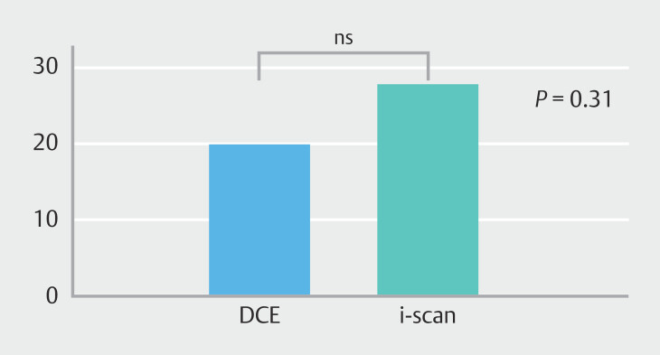
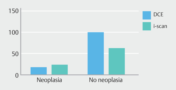
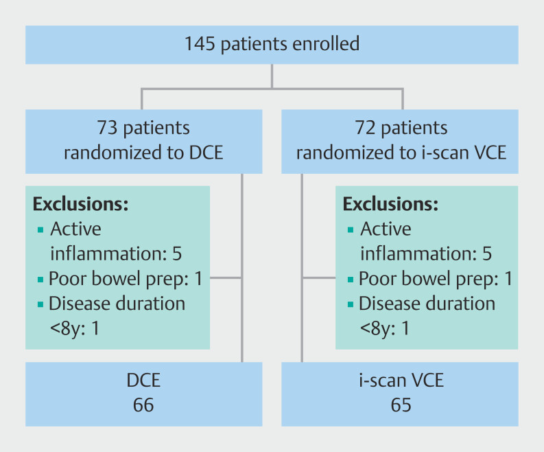
Background and study aims Long-standing ulcerative colitis (UC) is associated with an increased risk of developing colorectal neoplasia. Both dye-based chromoendoscopy (DCE) and virtual chromoendoscopy (VCE) increase detection of neoplastic lesions. In this prospective randomized controlled trial (RCT), we compared the neoplasia detection rate between DCE and i-scan VCE in patients with long-standing UC. Patient and methods In four European hospitals, 131 patients with long-standing UC (disease duration > 8 years) were randomized to either DCE with methylene blue 0.1% (n = 66) or i-scan VCE (n = 65). All procedures were performed by trained endoscopists. Biopsies were taken from all visible lesions and the surrounding mucosa. Results The mean number of neoplastic lesions detected per colonoscopy was not significantly different between DCE (0.27) and i-scan VCE (0.37) ( P = 0.41). Similarly, there was no significant difference in neoplasia detection rate between DCE (19.7%) and VCE (27.7%) (odds ratio0.64, 95% confidence interval 0.28-1.50, P = 0.31). However, the per lesion neoplasia detection rate was significantly higher with i-scan VCE compared to DCE (27.6% vs 15.3%, P = 0.036). Both withdrawal and total procedure time were on average 10.0 and 9.9 minutes shorter using i-scan VCE (both P < 0.001). Conclusions This multicenter, prospective RCT showed no significant difference in neoplasia detection between DCE and i-scan VCE in long-standing UC. However, use of i-scan VCE was associated with a lower false-positive rate and a significantly shorter procedure time compared with DCE. I-scan VCE, therefore, could be a valid replacement for DCE in UC surveillance colonoscopies.


 分享
分享
 求助内容:
求助内容: 应助结果提醒方式:
 扫码关注我们
