{"title":"视觉诱发电位对轻度颅脑损伤运动员大细胞和旁细胞通路的影响。","authors":"Mark H Myers, Nidhish Kalyanakumar, Paul Harris","doi":"10.1177/26331055241303165","DOIUrl":null,"url":null,"abstract":"<p><strong>Background: </strong>The objective of this study is to examine magnocellular and parvocellular pathways differentiation based on checkerboard spatial frequency stimulation between normal and visually impaired individuals from athletes with mild traumatic brain injury.</p><p><strong>Purpose: </strong>Athletes who exhibited photophobia, and blurriness were subjected to 5 spatial frequency stimuli presented to the left and right eye, and both eyes simultaneously to determine the type of receptive field loss deprecation based on sports-related brain trauma.</p><p><strong>Methods: </strong>Checkerboard stimulation enables the measurement between 2 visual processing pathways and enables the determination of the integrity of visual processing through visual evoked potentials (VEPs).</p><p><strong>Conclusion: </strong>The principal results reflect P1 responses demonstrated distinct changes in amplitude from mTBI (>5 µV) from normal cohorts concluding higher P1 amplitude of the VEP in mTBI cohorts had increased after injury. Latency in P1 was not as distinct as amplitude changes. Our major conclusion is that most of the mTBI cohort exhibited receptive field loss across all the patients appears to be magnocellular process deprecation due to frequent instances of 8 × 8 and 16 × 16 spatial frequencies input as it relates to amplitude and latency output.</p>","PeriodicalId":36527,"journal":{"name":"Neuroscience Insights","volume":"19 ","pages":"26331055241303165"},"PeriodicalIF":2.6000,"publicationDate":"2024-11-27","publicationTypes":"Journal Article","fieldsOfStudy":null,"isOpenAccess":false,"openAccessPdf":"https://www.ncbi.nlm.nih.gov/pmc/articles/PMC11603483/pdf/","citationCount":"0","resultStr":"{\"title\":\"Visual Evoked Potential Effects on Magnocellular and Parvocellular Pathways from Athletes After Mild Traumatic Brain Injuries.\",\"authors\":\"Mark H Myers, Nidhish Kalyanakumar, Paul Harris\",\"doi\":\"10.1177/26331055241303165\",\"DOIUrl\":null,\"url\":null,\"abstract\":\"<p><strong>Background: </strong>The objective of this study is to examine magnocellular and parvocellular pathways differentiation based on checkerboard spatial frequency stimulation between normal and visually impaired individuals from athletes with mild traumatic brain injury.</p><p><strong>Purpose: </strong>Athletes who exhibited photophobia, and blurriness were subjected to 5 spatial frequency stimuli presented to the left and right eye, and both eyes simultaneously to determine the type of receptive field loss deprecation based on sports-related brain trauma.</p><p><strong>Methods: </strong>Checkerboard stimulation enables the measurement between 2 visual processing pathways and enables the determination of the integrity of visual processing through visual evoked potentials (VEPs).</p><p><strong>Conclusion: </strong>The principal results reflect P1 responses demonstrated distinct changes in amplitude from mTBI (>5 µV) from normal cohorts concluding higher P1 amplitude of the VEP in mTBI cohorts had increased after injury. Latency in P1 was not as distinct as amplitude changes. Our major conclusion is that most of the mTBI cohort exhibited receptive field loss across all the patients appears to be magnocellular process deprecation due to frequent instances of 8 × 8 and 16 × 16 spatial frequencies input as it relates to amplitude and latency output.</p>\",\"PeriodicalId\":36527,\"journal\":{\"name\":\"Neuroscience Insights\",\"volume\":\"19 \",\"pages\":\"26331055241303165\"},\"PeriodicalIF\":2.6000,\"publicationDate\":\"2024-11-27\",\"publicationTypes\":\"Journal Article\",\"fieldsOfStudy\":null,\"isOpenAccess\":false,\"openAccessPdf\":\"https://www.ncbi.nlm.nih.gov/pmc/articles/PMC11603483/pdf/\",\"citationCount\":\"0\",\"resultStr\":null,\"platform\":\"Semanticscholar\",\"paperid\":null,\"PeriodicalName\":\"Neuroscience Insights\",\"FirstCategoryId\":\"1085\",\"ListUrlMain\":\"https://doi.org/10.1177/26331055241303165\",\"RegionNum\":0,\"RegionCategory\":null,\"ArticlePicture\":[],\"TitleCN\":null,\"AbstractTextCN\":null,\"PMCID\":null,\"EPubDate\":\"2024/1/1 0:00:00\",\"PubModel\":\"eCollection\",\"JCR\":\"Q2\",\"JCRName\":\"NEUROSCIENCES\",\"Score\":null,\"Total\":0}","platform":"Semanticscholar","paperid":null,"PeriodicalName":"Neuroscience Insights","FirstCategoryId":"1085","ListUrlMain":"https://doi.org/10.1177/26331055241303165","RegionNum":0,"RegionCategory":null,"ArticlePicture":[],"TitleCN":null,"AbstractTextCN":null,"PMCID":null,"EPubDate":"2024/1/1 0:00:00","PubModel":"eCollection","JCR":"Q2","JCRName":"NEUROSCIENCES","Score":null,"Total":0}
Visual Evoked Potential Effects on Magnocellular and Parvocellular Pathways from Athletes After Mild Traumatic Brain Injuries.
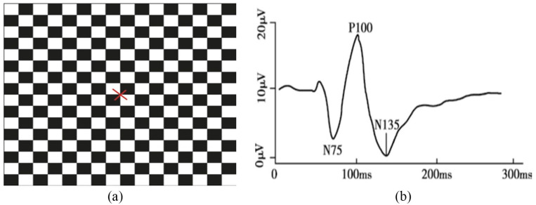
Background: The objective of this study is to examine magnocellular and parvocellular pathways differentiation based on checkerboard spatial frequency stimulation between normal and visually impaired individuals from athletes with mild traumatic brain injury.
Purpose: Athletes who exhibited photophobia, and blurriness were subjected to 5 spatial frequency stimuli presented to the left and right eye, and both eyes simultaneously to determine the type of receptive field loss deprecation based on sports-related brain trauma.
Methods: Checkerboard stimulation enables the measurement between 2 visual processing pathways and enables the determination of the integrity of visual processing through visual evoked potentials (VEPs).
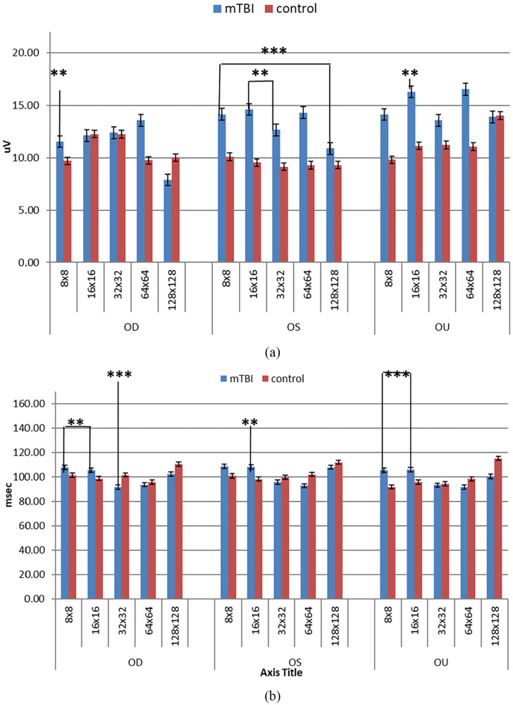
Conclusion: The principal results reflect P1 responses demonstrated distinct changes in amplitude from mTBI (>5 µV) from normal cohorts concluding higher P1 amplitude of the VEP in mTBI cohorts had increased after injury. Latency in P1 was not as distinct as amplitude changes. Our major conclusion is that most of the mTBI cohort exhibited receptive field loss across all the patients appears to be magnocellular process deprecation due to frequent instances of 8 × 8 and 16 × 16 spatial frequencies input as it relates to amplitude and latency output.


 分享
分享
 求助内容:
求助内容: 应助结果提醒方式:
 扫码关注我们
