{"title":"胆道癌合并先天性胆道扩张行胰十二指肠切除术及活体肝移植1例。","authors":"Tsuyoshi Shimamura, Masaaki Watanabe, Yasuyuki Koshizuka, Ryoichi Goto, Norio Kawamura, Tatsuya Orimo, Hirofumi Kamachi, Toshiya Kamiyama, Tomoko Mitsuhashi, Taizo Hibi, Akinobu Taketomi","doi":"10.1186/s40792-024-02068-5","DOIUrl":null,"url":null,"abstract":"<p><strong>Background: </strong>In patients with pancreaticobiliary maljunction complicated by congenital biliary dilatation, the pancreatic enzyme flows back into the bile, leading to bile duct carcinogenesis. Although the biliary tract resection and reconstruction is well documented to decrease the rate of malignancy, cancer occurrence has been reported in the residual intrahepatic or intrapancreatic bile duct, even after resection. We report a case of multiple biliary tract cancers in the liver complicated by congenital biliary dilatation, whose tumor lesions were resected en bloc without disconnecting the biliary tract by simultaneous pancreatoduodenectomy and living donor liver transplantation.</p><p><strong>Case presentation: </strong>A 27-year-old woman presented with epigastric discomfort. Examination indicated multiple biliary tract cancers complicated by congenital biliary dilatation. Computed tomography scan revealed three papillary tumors in the right hepatic duct with increased <sup>18</sup>F-FDG accumulation on positron emission tomography. Contrast-enhanced ultrasound revealed another lesion in the left hepatic duct. Adenocarcinoma cells were detected using bile and choledochal brush cytology. Tumors resection by right lobectomy or trisegmentectomy of the liver and extrahepatic bile duct resection indicated a high risk of postoperative liver failure; the residual liver volumes were calculated only 277 ml or 176 ml, respectively. In addition, tumor recurrence owing to bile leakage during the surgery and carcinogenesis from the remaining bile duct were concerned. Pancreatoduodenectomy was performed without disconnecting the biliary tract, and the tumors were resected en bloc with the whole liver. The left lobe liver graft from the husband was then transplanted. After 5 years of adjuvant treatment with tegafur/gimeracil/oteracil potassium, she remained in remission eight and half years after the surgery.</p><p><strong>Conclusions: </strong>Given the mechanism and development of cancer in the congenital biliary dilatation, simultaneous pancreatoduodenectomy and liver transplantation may be considered, especially in the case of young patients.</p>","PeriodicalId":22096,"journal":{"name":"Surgical Case Reports","volume":"10 1","pages":"274"},"PeriodicalIF":0.7000,"publicationDate":"2024-12-04","publicationTypes":"Journal Article","fieldsOfStudy":null,"isOpenAccess":false,"openAccessPdf":"https://www.ncbi.nlm.nih.gov/pmc/articles/PMC11618261/pdf/","citationCount":"0","resultStr":"{\"title\":\"A case of simultaneous pancreatoduodenectomy and living donor liver transplantation for biliary cancer complicated with congenital biliary dilatation.\",\"authors\":\"Tsuyoshi Shimamura, Masaaki Watanabe, Yasuyuki Koshizuka, Ryoichi Goto, Norio Kawamura, Tatsuya Orimo, Hirofumi Kamachi, Toshiya Kamiyama, Tomoko Mitsuhashi, Taizo Hibi, Akinobu Taketomi\",\"doi\":\"10.1186/s40792-024-02068-5\",\"DOIUrl\":null,\"url\":null,\"abstract\":\"<p><strong>Background: </strong>In patients with pancreaticobiliary maljunction complicated by congenital biliary dilatation, the pancreatic enzyme flows back into the bile, leading to bile duct carcinogenesis. Although the biliary tract resection and reconstruction is well documented to decrease the rate of malignancy, cancer occurrence has been reported in the residual intrahepatic or intrapancreatic bile duct, even after resection. We report a case of multiple biliary tract cancers in the liver complicated by congenital biliary dilatation, whose tumor lesions were resected en bloc without disconnecting the biliary tract by simultaneous pancreatoduodenectomy and living donor liver transplantation.</p><p><strong>Case presentation: </strong>A 27-year-old woman presented with epigastric discomfort. Examination indicated multiple biliary tract cancers complicated by congenital biliary dilatation. Computed tomography scan revealed three papillary tumors in the right hepatic duct with increased <sup>18</sup>F-FDG accumulation on positron emission tomography. Contrast-enhanced ultrasound revealed another lesion in the left hepatic duct. Adenocarcinoma cells were detected using bile and choledochal brush cytology. Tumors resection by right lobectomy or trisegmentectomy of the liver and extrahepatic bile duct resection indicated a high risk of postoperative liver failure; the residual liver volumes were calculated only 277 ml or 176 ml, respectively. In addition, tumor recurrence owing to bile leakage during the surgery and carcinogenesis from the remaining bile duct were concerned. Pancreatoduodenectomy was performed without disconnecting the biliary tract, and the tumors were resected en bloc with the whole liver. The left lobe liver graft from the husband was then transplanted. After 5 years of adjuvant treatment with tegafur/gimeracil/oteracil potassium, she remained in remission eight and half years after the surgery.</p><p><strong>Conclusions: </strong>Given the mechanism and development of cancer in the congenital biliary dilatation, simultaneous pancreatoduodenectomy and liver transplantation may be considered, especially in the case of young patients.</p>\",\"PeriodicalId\":22096,\"journal\":{\"name\":\"Surgical Case Reports\",\"volume\":\"10 1\",\"pages\":\"274\"},\"PeriodicalIF\":0.7000,\"publicationDate\":\"2024-12-04\",\"publicationTypes\":\"Journal Article\",\"fieldsOfStudy\":null,\"isOpenAccess\":false,\"openAccessPdf\":\"https://www.ncbi.nlm.nih.gov/pmc/articles/PMC11618261/pdf/\",\"citationCount\":\"0\",\"resultStr\":null,\"platform\":\"Semanticscholar\",\"paperid\":null,\"PeriodicalName\":\"Surgical Case Reports\",\"FirstCategoryId\":\"1085\",\"ListUrlMain\":\"https://doi.org/10.1186/s40792-024-02068-5\",\"RegionNum\":0,\"RegionCategory\":null,\"ArticlePicture\":[],\"TitleCN\":null,\"AbstractTextCN\":null,\"PMCID\":null,\"EPubDate\":\"\",\"PubModel\":\"\",\"JCR\":\"Q4\",\"JCRName\":\"SURGERY\",\"Score\":null,\"Total\":0}","platform":"Semanticscholar","paperid":null,"PeriodicalName":"Surgical Case Reports","FirstCategoryId":"1085","ListUrlMain":"https://doi.org/10.1186/s40792-024-02068-5","RegionNum":0,"RegionCategory":null,"ArticlePicture":[],"TitleCN":null,"AbstractTextCN":null,"PMCID":null,"EPubDate":"","PubModel":"","JCR":"Q4","JCRName":"SURGERY","Score":null,"Total":0}
A case of simultaneous pancreatoduodenectomy and living donor liver transplantation for biliary cancer complicated with congenital biliary dilatation.
Background: In patients with pancreaticobiliary maljunction complicated by congenital biliary dilatation, the pancreatic enzyme flows back into the bile, leading to bile duct carcinogenesis. Although the biliary tract resection and reconstruction is well documented to decrease the rate of malignancy, cancer occurrence has been reported in the residual intrahepatic or intrapancreatic bile duct, even after resection. We report a case of multiple biliary tract cancers in the liver complicated by congenital biliary dilatation, whose tumor lesions were resected en bloc without disconnecting the biliary tract by simultaneous pancreatoduodenectomy and living donor liver transplantation.
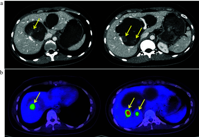
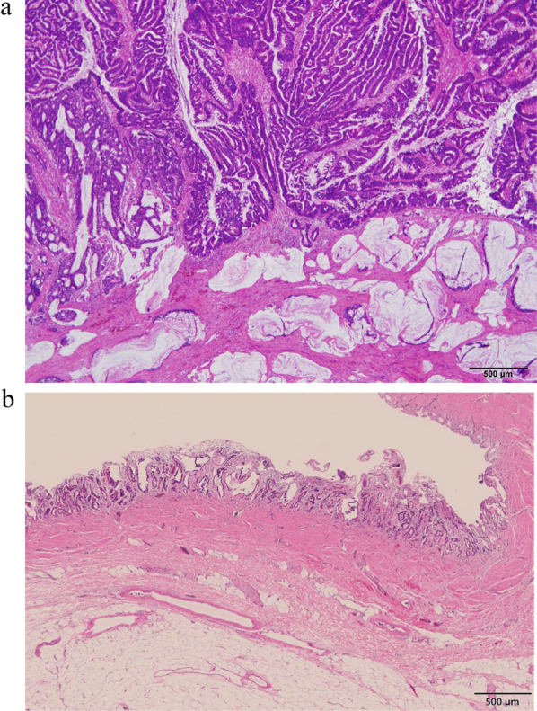
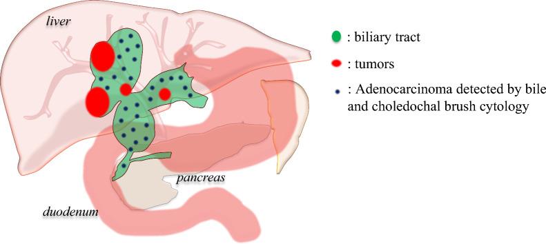
Case presentation: A 27-year-old woman presented with epigastric discomfort. Examination indicated multiple biliary tract cancers complicated by congenital biliary dilatation. Computed tomography scan revealed three papillary tumors in the right hepatic duct with increased 18F-FDG accumulation on positron emission tomography. Contrast-enhanced ultrasound revealed another lesion in the left hepatic duct. Adenocarcinoma cells were detected using bile and choledochal brush cytology. Tumors resection by right lobectomy or trisegmentectomy of the liver and extrahepatic bile duct resection indicated a high risk of postoperative liver failure; the residual liver volumes were calculated only 277 ml or 176 ml, respectively. In addition, tumor recurrence owing to bile leakage during the surgery and carcinogenesis from the remaining bile duct were concerned. Pancreatoduodenectomy was performed without disconnecting the biliary tract, and the tumors were resected en bloc with the whole liver. The left lobe liver graft from the husband was then transplanted. After 5 years of adjuvant treatment with tegafur/gimeracil/oteracil potassium, she remained in remission eight and half years after the surgery.
Conclusions: Given the mechanism and development of cancer in the congenital biliary dilatation, simultaneous pancreatoduodenectomy and liver transplantation may be considered, especially in the case of young patients.


 分享
分享
 求助内容:
求助内容: 应助结果提醒方式:
 扫码关注我们
