{"title":"急诊超声诊断小儿腹痛的罕见病因。","authors":"Courtney Owens, Lindsey Chaudoin","doi":"10.24908/pocus.v9i2.17451","DOIUrl":null,"url":null,"abstract":"<p><p>An 11-year-old postmenarchal female presented to the pediatric emergency department (ED) with 2 days of periumbilical and right lower abdominal pain. Radiology-performed ultrasound (RADUS) did not visualize the appendix, and there was a plan for surgical consultation to decide between serial abdominal exams versus computed tomography (CT) scan. While awaiting consultation and to help further narrow the differential diagnosis, the emergency provider performed a point of care ultrasound (POCUS) of the urinary tract. This revealed several anomalies including a solitary left kidney with hydronephrosis, and a well-circumscribed, fluid-filled structure with mixed echogenicity posterior to the bladder and inferior to the uterus. Given these findings on POCUS, further imaging was pursued, including a RADUS of the pelvis followed by a magnetic resonance imaging (MRI) of the abdomen. Further imaging ultimately diagnosed a bicornuate uterus, septate vagina with hematocolpos and solitary left kidney consistent with Obstructed Hemivagina and Ipsilateral Renal Anomaly (OHVIRA) syndrome. This case is an illustration of how POCUS is an invaluable tool to narrow the differential diagnosis and guide advanced imaging or consultation for both common and rare causes of pediatric abdominal pain.</p>","PeriodicalId":74470,"journal":{"name":"POCUS journal","volume":"9 2","pages":"32-34"},"PeriodicalIF":0.0000,"publicationDate":"2024-11-15","publicationTypes":"Journal Article","fieldsOfStudy":null,"isOpenAccess":false,"openAccessPdf":"https://www.ncbi.nlm.nih.gov/pmc/articles/PMC11614403/pdf/","citationCount":"0","resultStr":"{\"title\":\"Rare Cause of Pediatric Abdominal Pain Diagnosed on Point of Care Ultrasound (POCUS).\",\"authors\":\"Courtney Owens, Lindsey Chaudoin\",\"doi\":\"10.24908/pocus.v9i2.17451\",\"DOIUrl\":null,\"url\":null,\"abstract\":\"<p><p>An 11-year-old postmenarchal female presented to the pediatric emergency department (ED) with 2 days of periumbilical and right lower abdominal pain. Radiology-performed ultrasound (RADUS) did not visualize the appendix, and there was a plan for surgical consultation to decide between serial abdominal exams versus computed tomography (CT) scan. While awaiting consultation and to help further narrow the differential diagnosis, the emergency provider performed a point of care ultrasound (POCUS) of the urinary tract. This revealed several anomalies including a solitary left kidney with hydronephrosis, and a well-circumscribed, fluid-filled structure with mixed echogenicity posterior to the bladder and inferior to the uterus. Given these findings on POCUS, further imaging was pursued, including a RADUS of the pelvis followed by a magnetic resonance imaging (MRI) of the abdomen. Further imaging ultimately diagnosed a bicornuate uterus, septate vagina with hematocolpos and solitary left kidney consistent with Obstructed Hemivagina and Ipsilateral Renal Anomaly (OHVIRA) syndrome. This case is an illustration of how POCUS is an invaluable tool to narrow the differential diagnosis and guide advanced imaging or consultation for both common and rare causes of pediatric abdominal pain.</p>\",\"PeriodicalId\":74470,\"journal\":{\"name\":\"POCUS journal\",\"volume\":\"9 2\",\"pages\":\"32-34\"},\"PeriodicalIF\":0.0000,\"publicationDate\":\"2024-11-15\",\"publicationTypes\":\"Journal Article\",\"fieldsOfStudy\":null,\"isOpenAccess\":false,\"openAccessPdf\":\"https://www.ncbi.nlm.nih.gov/pmc/articles/PMC11614403/pdf/\",\"citationCount\":\"0\",\"resultStr\":null,\"platform\":\"Semanticscholar\",\"paperid\":null,\"PeriodicalName\":\"POCUS journal\",\"FirstCategoryId\":\"1085\",\"ListUrlMain\":\"https://doi.org/10.24908/pocus.v9i2.17451\",\"RegionNum\":0,\"RegionCategory\":null,\"ArticlePicture\":[],\"TitleCN\":null,\"AbstractTextCN\":null,\"PMCID\":null,\"EPubDate\":\"2024/1/1 0:00:00\",\"PubModel\":\"eCollection\",\"JCR\":\"\",\"JCRName\":\"\",\"Score\":null,\"Total\":0}","platform":"Semanticscholar","paperid":null,"PeriodicalName":"POCUS journal","FirstCategoryId":"1085","ListUrlMain":"https://doi.org/10.24908/pocus.v9i2.17451","RegionNum":0,"RegionCategory":null,"ArticlePicture":[],"TitleCN":null,"AbstractTextCN":null,"PMCID":null,"EPubDate":"2024/1/1 0:00:00","PubModel":"eCollection","JCR":"","JCRName":"","Score":null,"Total":0}
Rare Cause of Pediatric Abdominal Pain Diagnosed on Point of Care Ultrasound (POCUS).
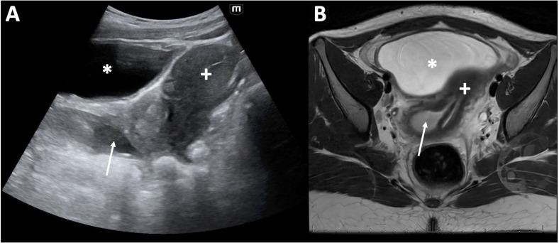
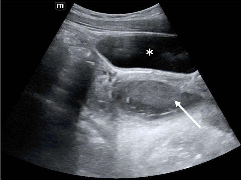
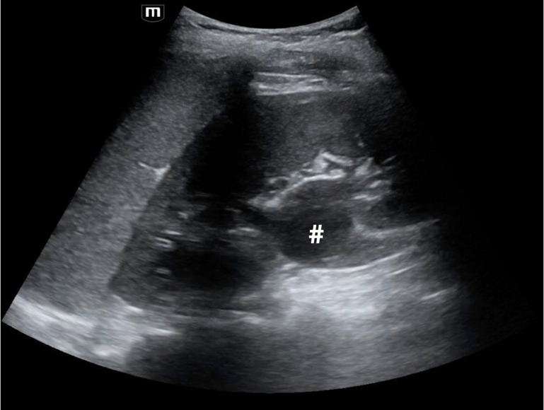
An 11-year-old postmenarchal female presented to the pediatric emergency department (ED) with 2 days of periumbilical and right lower abdominal pain. Radiology-performed ultrasound (RADUS) did not visualize the appendix, and there was a plan for surgical consultation to decide between serial abdominal exams versus computed tomography (CT) scan. While awaiting consultation and to help further narrow the differential diagnosis, the emergency provider performed a point of care ultrasound (POCUS) of the urinary tract. This revealed several anomalies including a solitary left kidney with hydronephrosis, and a well-circumscribed, fluid-filled structure with mixed echogenicity posterior to the bladder and inferior to the uterus. Given these findings on POCUS, further imaging was pursued, including a RADUS of the pelvis followed by a magnetic resonance imaging (MRI) of the abdomen. Further imaging ultimately diagnosed a bicornuate uterus, septate vagina with hematocolpos and solitary left kidney consistent with Obstructed Hemivagina and Ipsilateral Renal Anomaly (OHVIRA) syndrome. This case is an illustration of how POCUS is an invaluable tool to narrow the differential diagnosis and guide advanced imaging or consultation for both common and rare causes of pediatric abdominal pain.


 分享
分享

 求助内容:
求助内容: 应助结果提醒方式:
 扫码关注我们
