{"title":"圆锥角膜患者在巩膜晶状体佩戴期间Bruch膜开口随眼压变化的变化。","authors":"Langis Michaud, Steven Balourdet, Dan Samaha","doi":"10.1111/opo.13431","DOIUrl":null,"url":null,"abstract":"<p><strong>Purpose: </strong>The present study aimed to determine the potential impact of scleral lenses on intraocular pressure (IOP) by analysing the Bruch's membrane opening-minimum rim width (BMO-MRW) while the lenses are worn, in a population with keratoconus.</p><p><strong>Methods: </strong>Participants were required to have keratoconus and be successfully fitted with scleral lenses for at least 3 months. A new pair of optimised scleral lenses was provided before the study. During the first session, corneal biomechanics was assessed using an air tonometer, coupling Scheimpflug technology. Then, a scan of the optic nerve was carried out using optical coherence tomography (OCT) at 2 h intervals for 6 h. Particular attention was paid to identifying the BMO-MRW, which represents the smallest distance between the BMO and the internal limiting membrane. These tests were repeated, respecting the time at which the initial measurements were taken, while the scleral lens was worn. Results from only one eye were analysed.</p><p><strong>Results: </strong>A statistically significant change of 10.5 ± 3.6 μm (95% CI [241.3-473.1]; p = 0.02) in BMO-MRW was observed after 6 h of scleral lens wear, compared to measurements without lenses (4.8 ± 3.4 μm; 95% CI [285.1-439.7]; p = 0.18). The fluctuation was greater in participants with keratoconus than found in a previous study of regular corneas.</p><p><strong>Conclusion: </strong>BMO-MRW became significantly thinner after 6 h of scleral lens wear compared with measurements without lenses. These variations may be associated with a rise in IOP during lens wear. Close monitoring for optic head changes should be carried out for patients at risk. These results should be compared with future longer-term studies including a larger cohort of patients.</p>","PeriodicalId":19522,"journal":{"name":"Ophthalmic and Physiological Optics","volume":" ","pages":"405-415"},"PeriodicalIF":2.4000,"publicationDate":"2025-03-01","publicationTypes":"Journal Article","fieldsOfStudy":null,"isOpenAccess":false,"openAccessPdf":"https://www.ncbi.nlm.nih.gov/pmc/articles/PMC11823389/pdf/","citationCount":"0","resultStr":"{\"title\":\"Variation of Bruch's membrane opening in response to intraocular pressure change during scleral lens wear, in a population with keratoconus.\",\"authors\":\"Langis Michaud, Steven Balourdet, Dan Samaha\",\"doi\":\"10.1111/opo.13431\",\"DOIUrl\":null,\"url\":null,\"abstract\":\"<p><strong>Purpose: </strong>The present study aimed to determine the potential impact of scleral lenses on intraocular pressure (IOP) by analysing the Bruch's membrane opening-minimum rim width (BMO-MRW) while the lenses are worn, in a population with keratoconus.</p><p><strong>Methods: </strong>Participants were required to have keratoconus and be successfully fitted with scleral lenses for at least 3 months. A new pair of optimised scleral lenses was provided before the study. During the first session, corneal biomechanics was assessed using an air tonometer, coupling Scheimpflug technology. Then, a scan of the optic nerve was carried out using optical coherence tomography (OCT) at 2 h intervals for 6 h. Particular attention was paid to identifying the BMO-MRW, which represents the smallest distance between the BMO and the internal limiting membrane. These tests were repeated, respecting the time at which the initial measurements were taken, while the scleral lens was worn. Results from only one eye were analysed.</p><p><strong>Results: </strong>A statistically significant change of 10.5 ± 3.6 μm (95% CI [241.3-473.1]; p = 0.02) in BMO-MRW was observed after 6 h of scleral lens wear, compared to measurements without lenses (4.8 ± 3.4 μm; 95% CI [285.1-439.7]; p = 0.18). The fluctuation was greater in participants with keratoconus than found in a previous study of regular corneas.</p><p><strong>Conclusion: </strong>BMO-MRW became significantly thinner after 6 h of scleral lens wear compared with measurements without lenses. These variations may be associated with a rise in IOP during lens wear. Close monitoring for optic head changes should be carried out for patients at risk. These results should be compared with future longer-term studies including a larger cohort of patients.</p>\",\"PeriodicalId\":19522,\"journal\":{\"name\":\"Ophthalmic and Physiological Optics\",\"volume\":\" \",\"pages\":\"405-415\"},\"PeriodicalIF\":2.4000,\"publicationDate\":\"2025-03-01\",\"publicationTypes\":\"Journal Article\",\"fieldsOfStudy\":null,\"isOpenAccess\":false,\"openAccessPdf\":\"https://www.ncbi.nlm.nih.gov/pmc/articles/PMC11823389/pdf/\",\"citationCount\":\"0\",\"resultStr\":null,\"platform\":\"Semanticscholar\",\"paperid\":null,\"PeriodicalName\":\"Ophthalmic and Physiological Optics\",\"FirstCategoryId\":\"3\",\"ListUrlMain\":\"https://doi.org/10.1111/opo.13431\",\"RegionNum\":3,\"RegionCategory\":\"医学\",\"ArticlePicture\":[],\"TitleCN\":null,\"AbstractTextCN\":null,\"PMCID\":null,\"EPubDate\":\"2024/12/6 0:00:00\",\"PubModel\":\"Epub\",\"JCR\":\"Q1\",\"JCRName\":\"OPHTHALMOLOGY\",\"Score\":null,\"Total\":0}","platform":"Semanticscholar","paperid":null,"PeriodicalName":"Ophthalmic and Physiological Optics","FirstCategoryId":"3","ListUrlMain":"https://doi.org/10.1111/opo.13431","RegionNum":3,"RegionCategory":"医学","ArticlePicture":[],"TitleCN":null,"AbstractTextCN":null,"PMCID":null,"EPubDate":"2024/12/6 0:00:00","PubModel":"Epub","JCR":"Q1","JCRName":"OPHTHALMOLOGY","Score":null,"Total":0}
引用次数: 0
摘要
目的:本研究旨在通过分析角膜圆锥患者佩戴巩膜晶状体时的Bruch膜开口-最小边缘宽度(BMO-MRW)来确定巩膜晶状体对眼压(IOP)的潜在影响。方法:参与者被要求患有圆锥角膜并成功植入巩膜晶体至少3个月。在研究前提供了一对新的优化巩膜镜片。在第一次会议中,使用空气血压计结合Scheimpflug技术评估角膜生物力学。然后,使用光学相干断层扫描(OCT)对视神经进行扫描,间隔2小时,持续6小时。特别注意的是识别BMO- mrw,它代表BMO和内限制膜之间的最小距离。在佩戴巩膜晶状体的同时,根据最初测量的时间,重复进行这些测试。只分析一只眼睛的结果。结果:差异有统计学意义,为10.5±3.6 μm (95% CI [241.3 ~ 473.1];p = 0.02),与未佩戴巩膜镜片(4.8±3.4 μm;95% ci [285.1-439.7];p = 0.18)。圆锥角膜患者的波动比之前对普通角膜的研究发现的要大。结论:在巩膜晶状体佩戴6小时后,BMO-MRW与未佩戴晶状体的测量结果相比明显变薄。这些变化可能与晶状体佩戴期间IOP升高有关。对于有危险的患者,应密切监测视头的变化。这些结果应该与未来的长期研究进行比较,包括更大的患者队列。
Variation of Bruch's membrane opening in response to intraocular pressure change during scleral lens wear, in a population with keratoconus.
Purpose: The present study aimed to determine the potential impact of scleral lenses on intraocular pressure (IOP) by analysing the Bruch's membrane opening-minimum rim width (BMO-MRW) while the lenses are worn, in a population with keratoconus.
Methods: Participants were required to have keratoconus and be successfully fitted with scleral lenses for at least 3 months. A new pair of optimised scleral lenses was provided before the study. During the first session, corneal biomechanics was assessed using an air tonometer, coupling Scheimpflug technology. Then, a scan of the optic nerve was carried out using optical coherence tomography (OCT) at 2 h intervals for 6 h. Particular attention was paid to identifying the BMO-MRW, which represents the smallest distance between the BMO and the internal limiting membrane. These tests were repeated, respecting the time at which the initial measurements were taken, while the scleral lens was worn. Results from only one eye were analysed.
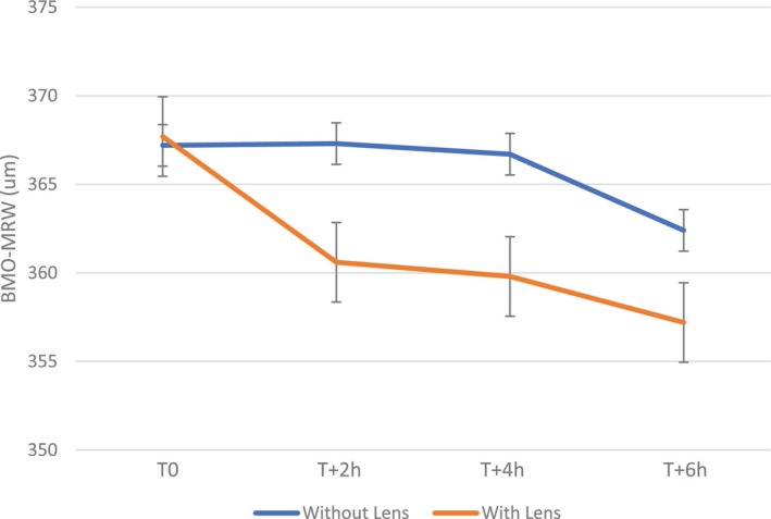
Results: A statistically significant change of 10.5 ± 3.6 μm (95% CI [241.3-473.1]; p = 0.02) in BMO-MRW was observed after 6 h of scleral lens wear, compared to measurements without lenses (4.8 ± 3.4 μm; 95% CI [285.1-439.7]; p = 0.18). The fluctuation was greater in participants with keratoconus than found in a previous study of regular corneas.
Conclusion: BMO-MRW became significantly thinner after 6 h of scleral lens wear compared with measurements without lenses. These variations may be associated with a rise in IOP during lens wear. Close monitoring for optic head changes should be carried out for patients at risk. These results should be compared with future longer-term studies including a larger cohort of patients.
期刊介绍:
Ophthalmic & Physiological Optics, first published in 1925, is a leading international interdisciplinary journal that addresses basic and applied questions pertinent to contemporary research in vision science and optometry.
OPO publishes original research papers, technical notes, reviews and letters and will interest researchers, educators and clinicians concerned with the development, use and restoration of vision.


 分享
分享
 求助内容:
求助内容: 应助结果提醒方式:
 扫码关注我们
