Elmira Khiabani, Anna C J Kalisvaart, Cassandra M Wilkinson, Peter L Hurd, Brian H Buck, Frederick Colbourne
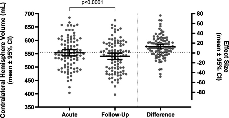
{"title":"评价mono - kellie学说:脑出血患者对侧半球萎缩。","authors":"Elmira Khiabani, Anna C J Kalisvaart, Cassandra M Wilkinson, Peter L Hurd, Brian H Buck, Frederick Colbourne","doi":"10.1007/s12975-024-01316-y","DOIUrl":null,"url":null,"abstract":"<p><p>Intracerebral hemorrhage (ICH) along with aggravating factors, such as edema, can raise intracranial pressure (ICP) to pathological levels. Diversion of some cerebrospinal fluid (CSF) and venous blood out of the cranium can limit ICP rises while maintaining cerebral perfusion pressure. Brain tissue itself is widely considered immutable in volume but prone to distortion (e.g., midline shift). However, distal brain regions shrink acutely following ICH in rodents. Tissue contraction arises from cell shrinkage and increased packing density. This \"tissue compliance\" is hypothesized to be an additional mechanism to limit ICP rises. Here, we examined whether and by how much parenchyma volume reduction occurs in ICH patients. We conducted a retrospective analysis on computed tomography (CT) scans of 96 ICH patients (average age 63.8 years old, 55% male) with an average hematoma volume of 32.4 and 35.3 mL at the first and second scan (separated by ~ 23 h), respectively. Hematoma growth (any absolute increase) occurred in 44% of patients, with a minimal but significant growth of the hematoma of 2.9 mL on average across all patients (p = 0.028). As hypothesized, the contralateral hemisphere volume was significantly reduced by 12.7 mL (p < 0.0001) between scans. This was unrelated to midline shift (R<sup>2</sup> = 0.012, p = 0.21), which averaged 2.3 mm. These findings suggest that distal parenchymal shrinkage may be a major compliance mechanism after ICH; the implications for ICP and brain function merit further study.</p>","PeriodicalId":23237,"journal":{"name":"Translational Stroke Research","volume":" ","pages":"1447-1451"},"PeriodicalIF":4.3000,"publicationDate":"2025-10-01","publicationTypes":"Journal Article","fieldsOfStudy":null,"isOpenAccess":false,"openAccessPdf":"https://www.ncbi.nlm.nih.gov/pmc/articles/PMC12391140/pdf/","citationCount":"0","resultStr":"{\"title\":\"Evaluating the Monro-Kellie Doctrine: Contralateral Hemisphere Shrinkage in Intracerebral Hemorrhage Patients.\",\"authors\":\"Elmira Khiabani, Anna C J Kalisvaart, Cassandra M Wilkinson, Peter L Hurd, Brian H Buck, Frederick Colbourne\",\"doi\":\"10.1007/s12975-024-01316-y\",\"DOIUrl\":null,\"url\":null,\"abstract\":\"<p><p>Intracerebral hemorrhage (ICH) along with aggravating factors, such as edema, can raise intracranial pressure (ICP) to pathological levels. Diversion of some cerebrospinal fluid (CSF) and venous blood out of the cranium can limit ICP rises while maintaining cerebral perfusion pressure. Brain tissue itself is widely considered immutable in volume but prone to distortion (e.g., midline shift). However, distal brain regions shrink acutely following ICH in rodents. Tissue contraction arises from cell shrinkage and increased packing density. This \\\"tissue compliance\\\" is hypothesized to be an additional mechanism to limit ICP rises. Here, we examined whether and by how much parenchyma volume reduction occurs in ICH patients. We conducted a retrospective analysis on computed tomography (CT) scans of 96 ICH patients (average age 63.8 years old, 55% male) with an average hematoma volume of 32.4 and 35.3 mL at the first and second scan (separated by ~ 23 h), respectively. Hematoma growth (any absolute increase) occurred in 44% of patients, with a minimal but significant growth of the hematoma of 2.9 mL on average across all patients (p = 0.028). As hypothesized, the contralateral hemisphere volume was significantly reduced by 12.7 mL (p < 0.0001) between scans. This was unrelated to midline shift (R<sup>2</sup> = 0.012, p = 0.21), which averaged 2.3 mm. These findings suggest that distal parenchymal shrinkage may be a major compliance mechanism after ICH; the implications for ICP and brain function merit further study.</p>\",\"PeriodicalId\":23237,\"journal\":{\"name\":\"Translational Stroke Research\",\"volume\":\" \",\"pages\":\"1447-1451\"},\"PeriodicalIF\":4.3000,\"publicationDate\":\"2025-10-01\",\"publicationTypes\":\"Journal Article\",\"fieldsOfStudy\":null,\"isOpenAccess\":false,\"openAccessPdf\":\"https://www.ncbi.nlm.nih.gov/pmc/articles/PMC12391140/pdf/\",\"citationCount\":\"0\",\"resultStr\":null,\"platform\":\"Semanticscholar\",\"paperid\":null,\"PeriodicalName\":\"Translational Stroke Research\",\"FirstCategoryId\":\"3\",\"ListUrlMain\":\"https://doi.org/10.1007/s12975-024-01316-y\",\"RegionNum\":2,\"RegionCategory\":\"医学\",\"ArticlePicture\":[],\"TitleCN\":null,\"AbstractTextCN\":null,\"PMCID\":null,\"EPubDate\":\"2024/12/11 0:00:00\",\"PubModel\":\"Epub\",\"JCR\":\"Q1\",\"JCRName\":\"CLINICAL NEUROLOGY\",\"Score\":null,\"Total\":0}","platform":"Semanticscholar","paperid":null,"PeriodicalName":"Translational Stroke Research","FirstCategoryId":"3","ListUrlMain":"https://doi.org/10.1007/s12975-024-01316-y","RegionNum":2,"RegionCategory":"医学","ArticlePicture":[],"TitleCN":null,"AbstractTextCN":null,"PMCID":null,"EPubDate":"2024/12/11 0:00:00","PubModel":"Epub","JCR":"Q1","JCRName":"CLINICAL NEUROLOGY","Score":null,"Total":0}
引用次数: 0
摘要
脑出血(ICH)伴加重因素,如水肿,可使颅内压(ICP)升高到病理水平。转移部分脑脊液和静脉血出颅,可在维持脑灌注压的同时限制颅内压升高。脑组织本身被广泛认为在体积上是不变的,但容易变形(例如,中线移位)。然而,在啮齿动物脑出血后,远端脑区急剧萎缩。组织收缩是由细胞收缩和堆积密度增加引起的。这种“组织顺应性”被假设为限制ICP升高的附加机制。在这里,我们研究了脑出血患者是否发生实质体积减少以及减少多少。我们回顾性分析96例脑出血患者(平均年龄63.8岁,男性55%)的CT扫描,第一次和第二次扫描(间隔约23 h)平均血肿量分别为32.4和35.3 mL。44%的患者出现血肿增长(任何绝对增长),所有患者的血肿平均增长2.9 mL,最小但显著(p = 0.028)。正如假设的那样,对侧半球体积显著减少12.7 mL (p 2 = 0.012, p = 0.21),平均减少2.3 mm。这些结果表明,远端实质收缩可能是脑出血后的主要顺应性机制;对颅内压和脑功能的影响值得进一步研究。
Evaluating the Monro-Kellie Doctrine: Contralateral Hemisphere Shrinkage in Intracerebral Hemorrhage Patients.
Intracerebral hemorrhage (ICH) along with aggravating factors, such as edema, can raise intracranial pressure (ICP) to pathological levels. Diversion of some cerebrospinal fluid (CSF) and venous blood out of the cranium can limit ICP rises while maintaining cerebral perfusion pressure. Brain tissue itself is widely considered immutable in volume but prone to distortion (e.g., midline shift). However, distal brain regions shrink acutely following ICH in rodents. Tissue contraction arises from cell shrinkage and increased packing density. This "tissue compliance" is hypothesized to be an additional mechanism to limit ICP rises. Here, we examined whether and by how much parenchyma volume reduction occurs in ICH patients. We conducted a retrospective analysis on computed tomography (CT) scans of 96 ICH patients (average age 63.8 years old, 55% male) with an average hematoma volume of 32.4 and 35.3 mL at the first and second scan (separated by ~ 23 h), respectively. Hematoma growth (any absolute increase) occurred in 44% of patients, with a minimal but significant growth of the hematoma of 2.9 mL on average across all patients (p = 0.028). As hypothesized, the contralateral hemisphere volume was significantly reduced by 12.7 mL (p < 0.0001) between scans. This was unrelated to midline shift (R2 = 0.012, p = 0.21), which averaged 2.3 mm. These findings suggest that distal parenchymal shrinkage may be a major compliance mechanism after ICH; the implications for ICP and brain function merit further study.
期刊介绍:
Translational Stroke Research covers basic, translational, and clinical studies. The Journal emphasizes novel approaches to help both to understand clinical phenomenon through basic science tools, and to translate basic science discoveries into the development of new strategies for the prevention, assessment, treatment, and enhancement of central nervous system repair after stroke and other forms of neurotrauma.
Translational Stroke Research focuses on translational research and is relevant to both basic scientists and physicians, including but not restricted to neuroscientists, vascular biologists, neurologists, neuroimagers, and neurosurgeons.


 分享
分享
 求助内容:
求助内容: 应助结果提醒方式:
 扫码关注我们
