Patrick Klepits, Karl Koschutnig, Thomas Zussner, Andreas Fink
{"title":"中等强度跑步干预后海马体积和情感功能的变化。","authors":"Patrick Klepits, Karl Koschutnig, Thomas Zussner, Andreas Fink","doi":"10.1007/s00429-024-02885-2","DOIUrl":null,"url":null,"abstract":"<p><p>This study examined the effects of a moderately intense seven-week running intervention on the hippocampal volume and depressive symptoms of young men (20-31 years of age) from the general population (N = 21). A within-subjects-design involving a two-week baseline period before the running intervention, and two subsequent intervention cycles was applied. At four time points of assessment (t<sub>1</sub>: start of the study; t<sub>2</sub>: end of baseline period/start of the intervention; t<sub>3</sub>: end of the first intervention cycle; t<sub>4</sub>: end of the 2nd intervention cycle/study end) magnetic resonance imaging was performed and symptoms related to depression were assessed employing the Center for Epidemiological Studies Depression (CES-D) Scale. The intervention resulted in a significant increase in the estimated maximum oxygen uptake (VO<sub>2</sub>max), measured with a standardized walking test (average increase from 42.07 ml*kg<sup>- 1</sup>*min<sup>- 1</sup> to 46.07 ml*kg<sup>- 1</sup>*min<sup>- 1</sup>). The CES-D scores decreased significantly over the course of the running intervention (average decrease from 12.76 to 10.48 on a 20-point scale). Significant volumetric increases in the hippocampus were found, most notably after the first intervention cycle in the left (average increase from 613.41 mm³ to 620.55 mm³) and right hippocampal tail (average increase from 629.77 mm³ to 638.17 mm³). These findings provide new evidence regarding the temporal dynamics of hippocampal changes following engagement in physical activity.</p>","PeriodicalId":9145,"journal":{"name":"Brain Structure & Function","volume":"230 1","pages":"2"},"PeriodicalIF":3.0000,"publicationDate":"2024-12-13","publicationTypes":"Journal Article","fieldsOfStudy":null,"isOpenAccess":false,"openAccessPdf":"https://www.ncbi.nlm.nih.gov/pmc/articles/PMC11645311/pdf/","citationCount":"0","resultStr":"{\"title\":\"Changes in hippocampal volume and affective functioning after a moderate intensity running intervention.\",\"authors\":\"Patrick Klepits, Karl Koschutnig, Thomas Zussner, Andreas Fink\",\"doi\":\"10.1007/s00429-024-02885-2\",\"DOIUrl\":null,\"url\":null,\"abstract\":\"<p><p>This study examined the effects of a moderately intense seven-week running intervention on the hippocampal volume and depressive symptoms of young men (20-31 years of age) from the general population (N = 21). A within-subjects-design involving a two-week baseline period before the running intervention, and two subsequent intervention cycles was applied. At four time points of assessment (t<sub>1</sub>: start of the study; t<sub>2</sub>: end of baseline period/start of the intervention; t<sub>3</sub>: end of the first intervention cycle; t<sub>4</sub>: end of the 2nd intervention cycle/study end) magnetic resonance imaging was performed and symptoms related to depression were assessed employing the Center for Epidemiological Studies Depression (CES-D) Scale. The intervention resulted in a significant increase in the estimated maximum oxygen uptake (VO<sub>2</sub>max), measured with a standardized walking test (average increase from 42.07 ml*kg<sup>- 1</sup>*min<sup>- 1</sup> to 46.07 ml*kg<sup>- 1</sup>*min<sup>- 1</sup>). The CES-D scores decreased significantly over the course of the running intervention (average decrease from 12.76 to 10.48 on a 20-point scale). Significant volumetric increases in the hippocampus were found, most notably after the first intervention cycle in the left (average increase from 613.41 mm³ to 620.55 mm³) and right hippocampal tail (average increase from 629.77 mm³ to 638.17 mm³). These findings provide new evidence regarding the temporal dynamics of hippocampal changes following engagement in physical activity.</p>\",\"PeriodicalId\":9145,\"journal\":{\"name\":\"Brain Structure & Function\",\"volume\":\"230 1\",\"pages\":\"2\"},\"PeriodicalIF\":3.0000,\"publicationDate\":\"2024-12-13\",\"publicationTypes\":\"Journal Article\",\"fieldsOfStudy\":null,\"isOpenAccess\":false,\"openAccessPdf\":\"https://www.ncbi.nlm.nih.gov/pmc/articles/PMC11645311/pdf/\",\"citationCount\":\"0\",\"resultStr\":null,\"platform\":\"Semanticscholar\",\"paperid\":null,\"PeriodicalName\":\"Brain Structure & Function\",\"FirstCategoryId\":\"3\",\"ListUrlMain\":\"https://doi.org/10.1007/s00429-024-02885-2\",\"RegionNum\":3,\"RegionCategory\":\"医学\",\"ArticlePicture\":[],\"TitleCN\":null,\"AbstractTextCN\":null,\"PMCID\":null,\"EPubDate\":\"\",\"PubModel\":\"\",\"JCR\":\"Q1\",\"JCRName\":\"ANATOMY & MORPHOLOGY\",\"Score\":null,\"Total\":0}","platform":"Semanticscholar","paperid":null,"PeriodicalName":"Brain Structure & Function","FirstCategoryId":"3","ListUrlMain":"https://doi.org/10.1007/s00429-024-02885-2","RegionNum":3,"RegionCategory":"医学","ArticlePicture":[],"TitleCN":null,"AbstractTextCN":null,"PMCID":null,"EPubDate":"","PubModel":"","JCR":"Q1","JCRName":"ANATOMY & MORPHOLOGY","Score":null,"Total":0}
Changes in hippocampal volume and affective functioning after a moderate intensity running intervention.
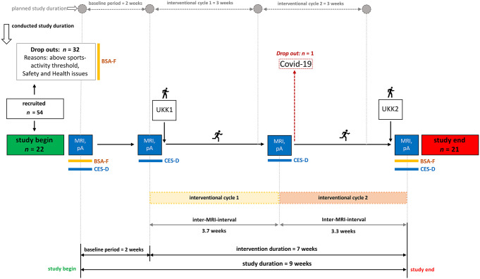
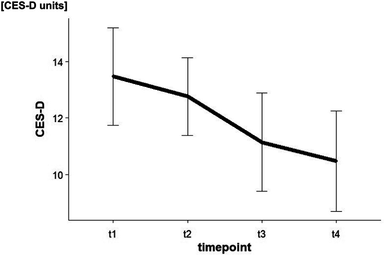
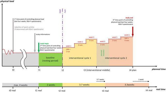
This study examined the effects of a moderately intense seven-week running intervention on the hippocampal volume and depressive symptoms of young men (20-31 years of age) from the general population (N = 21). A within-subjects-design involving a two-week baseline period before the running intervention, and two subsequent intervention cycles was applied. At four time points of assessment (t1: start of the study; t2: end of baseline period/start of the intervention; t3: end of the first intervention cycle; t4: end of the 2nd intervention cycle/study end) magnetic resonance imaging was performed and symptoms related to depression were assessed employing the Center for Epidemiological Studies Depression (CES-D) Scale. The intervention resulted in a significant increase in the estimated maximum oxygen uptake (VO2max), measured with a standardized walking test (average increase from 42.07 ml*kg- 1*min- 1 to 46.07 ml*kg- 1*min- 1). The CES-D scores decreased significantly over the course of the running intervention (average decrease from 12.76 to 10.48 on a 20-point scale). Significant volumetric increases in the hippocampus were found, most notably after the first intervention cycle in the left (average increase from 613.41 mm³ to 620.55 mm³) and right hippocampal tail (average increase from 629.77 mm³ to 638.17 mm³). These findings provide new evidence regarding the temporal dynamics of hippocampal changes following engagement in physical activity.
期刊介绍:
Brain Structure & Function publishes research that provides insight into brain structure−function relationships. Studies published here integrate data spanning from molecular, cellular, developmental, and systems architecture to the neuroanatomy of behavior and cognitive functions. Manuscripts with focus on the spinal cord or the peripheral nervous system are not accepted for publication. Manuscripts with focus on diseases, animal models of diseases, or disease-related mechanisms are only considered for publication, if the findings provide novel insight into the organization and mechanisms of normal brain structure and function.


 分享
分享
 求助内容:
求助内容: 应助结果提醒方式:
 扫码关注我们
