Adersh Stanly, Saumya Sara Sunny, Justin Benjamin, Hesarghatta Shyamasunde Asha, David Mathew, Junita Rachel John, Julie Hephzibah
{"title":"F18-FDG PET/CT在垂体摄取评价中的应用。","authors":"Adersh Stanly, Saumya Sara Sunny, Justin Benjamin, Hesarghatta Shyamasunde Asha, David Mathew, Junita Rachel John, Julie Hephzibah","doi":"10.1055/s-0044-1787967","DOIUrl":null,"url":null,"abstract":"<p><p><b>Introduction</b> Pituitary adenoma is the most common disease that affects the gland and may be classified as functional/nonsecretory tumors. Inflammatory/infective causes may also affect the pituitary gland. The 18F-fluorodeoxyglucose positron emission tomography/computed tomography (F18-FDG PET/CT) may have an incremental value in assessing these lesions and in determining their clinical significance. <b>Aim</b> This article assesses the utility of F18-FDG PET/CT in detecting and determining clinical profile of pituitary lesions with abnormal uptake. <b>Methodology</b> Retrospective analysis of all patients who underwent F18-FDG PET/CT from January 2015 to January 2023 was done. Those with abnormal pituitary uptake (standardized uptake value [SUV] > 2.5) were included in the study. SUV value along with relevant anatomical details, biochemical parameters, histopathological details, and follow-up imaging were analyzed. <b>Results</b> Among 15,085 studies, a total of 36 patients (21 males/15 females, average age 47.36 years, range: 17-75 years) with pituitary uptake (0.23%) were included. Out of 36 patients, causes are primary pituitary tumor (21/36, 58%), tubercular hypophysitis (3/36, 8%), lymphocytic hypophysitis (2/36, 6%), lymphomatous involvement (2/36, 6%), autoimmune hypophysitis (1/36, 3%), questionable significance/incidental (4/36, 11%), and metastasis (3/36, 8%)-one each from neuroendocrine tumor ileum, chondrosarcoma, and adenocarcinoma lung. There was no difference in the SUV range between the different etiologies. Among 21 patients with pituitary tumor, biochemical evaluation was done in 19 patients. Two patients were lost to follow-up and did not have biochemical evaluation. Among them, 8 underwent endoscopic transsphenoidal radical excision and 1 patient had PET-CT-guided stereotactic radiosurgery alone. In another 8 patients who had prior endoscopic transsphenoidal radical excision, uptake was noted as residual lesion on PET-CT. Of them, 3 underwent subtotal excision and 5 had PET-CT-guided stereotactic radiosurgery. Biopsy was done in 14 patients, of which 11 were macroadenoma and 3 were microadenoma. Overall, magnetic resonance imaging (MRI) brain was performed in 22 of them and the findings were concordant with F18-FDG PET/CT. <b>Conclusion</b> F18-FDG PET/CT is a useful modality in the evaluation of pituitary uptake. It has an incremental value along with MRI brain and biochemical parameters and is useful for follow-up. Due to its high diagnostic accuracy, it is particularly useful in those with suspected residual/recurrent adenomas.</p>","PeriodicalId":23742,"journal":{"name":"World Journal of Nuclear Medicine","volume":"23 4","pages":"234-241"},"PeriodicalIF":0.9000,"publicationDate":"2024-06-25","publicationTypes":"Journal Article","fieldsOfStudy":null,"isOpenAccess":false,"openAccessPdf":"https://www.ncbi.nlm.nih.gov/pmc/articles/PMC11637635/pdf/","citationCount":"0","resultStr":"{\"title\":\"Utility of F18-FDG PET/CT in the Evaluation of Pituitary Uptake.\",\"authors\":\"Adersh Stanly, Saumya Sara Sunny, Justin Benjamin, Hesarghatta Shyamasunde Asha, David Mathew, Junita Rachel John, Julie Hephzibah\",\"doi\":\"10.1055/s-0044-1787967\",\"DOIUrl\":null,\"url\":null,\"abstract\":\"<p><p><b>Introduction</b> Pituitary adenoma is the most common disease that affects the gland and may be classified as functional/nonsecretory tumors. Inflammatory/infective causes may also affect the pituitary gland. The 18F-fluorodeoxyglucose positron emission tomography/computed tomography (F18-FDG PET/CT) may have an incremental value in assessing these lesions and in determining their clinical significance. <b>Aim</b> This article assesses the utility of F18-FDG PET/CT in detecting and determining clinical profile of pituitary lesions with abnormal uptake. <b>Methodology</b> Retrospective analysis of all patients who underwent F18-FDG PET/CT from January 2015 to January 2023 was done. Those with abnormal pituitary uptake (standardized uptake value [SUV] > 2.5) were included in the study. SUV value along with relevant anatomical details, biochemical parameters, histopathological details, and follow-up imaging were analyzed. <b>Results</b> Among 15,085 studies, a total of 36 patients (21 males/15 females, average age 47.36 years, range: 17-75 years) with pituitary uptake (0.23%) were included. Out of 36 patients, causes are primary pituitary tumor (21/36, 58%), tubercular hypophysitis (3/36, 8%), lymphocytic hypophysitis (2/36, 6%), lymphomatous involvement (2/36, 6%), autoimmune hypophysitis (1/36, 3%), questionable significance/incidental (4/36, 11%), and metastasis (3/36, 8%)-one each from neuroendocrine tumor ileum, chondrosarcoma, and adenocarcinoma lung. There was no difference in the SUV range between the different etiologies. Among 21 patients with pituitary tumor, biochemical evaluation was done in 19 patients. Two patients were lost to follow-up and did not have biochemical evaluation. Among them, 8 underwent endoscopic transsphenoidal radical excision and 1 patient had PET-CT-guided stereotactic radiosurgery alone. In another 8 patients who had prior endoscopic transsphenoidal radical excision, uptake was noted as residual lesion on PET-CT. Of them, 3 underwent subtotal excision and 5 had PET-CT-guided stereotactic radiosurgery. Biopsy was done in 14 patients, of which 11 were macroadenoma and 3 were microadenoma. Overall, magnetic resonance imaging (MRI) brain was performed in 22 of them and the findings were concordant with F18-FDG PET/CT. <b>Conclusion</b> F18-FDG PET/CT is a useful modality in the evaluation of pituitary uptake. It has an incremental value along with MRI brain and biochemical parameters and is useful for follow-up. Due to its high diagnostic accuracy, it is particularly useful in those with suspected residual/recurrent adenomas.</p>\",\"PeriodicalId\":23742,\"journal\":{\"name\":\"World Journal of Nuclear Medicine\",\"volume\":\"23 4\",\"pages\":\"234-241\"},\"PeriodicalIF\":0.9000,\"publicationDate\":\"2024-06-25\",\"publicationTypes\":\"Journal Article\",\"fieldsOfStudy\":null,\"isOpenAccess\":false,\"openAccessPdf\":\"https://www.ncbi.nlm.nih.gov/pmc/articles/PMC11637635/pdf/\",\"citationCount\":\"0\",\"resultStr\":null,\"platform\":\"Semanticscholar\",\"paperid\":null,\"PeriodicalName\":\"World Journal of Nuclear Medicine\",\"FirstCategoryId\":\"1085\",\"ListUrlMain\":\"https://doi.org/10.1055/s-0044-1787967\",\"RegionNum\":0,\"RegionCategory\":null,\"ArticlePicture\":[],\"TitleCN\":null,\"AbstractTextCN\":null,\"PMCID\":null,\"EPubDate\":\"2024/12/1 0:00:00\",\"PubModel\":\"eCollection\",\"JCR\":\"Q4\",\"JCRName\":\"RADIOLOGY, NUCLEAR MEDICINE & MEDICAL IMAGING\",\"Score\":null,\"Total\":0}","platform":"Semanticscholar","paperid":null,"PeriodicalName":"World Journal of Nuclear Medicine","FirstCategoryId":"1085","ListUrlMain":"https://doi.org/10.1055/s-0044-1787967","RegionNum":0,"RegionCategory":null,"ArticlePicture":[],"TitleCN":null,"AbstractTextCN":null,"PMCID":null,"EPubDate":"2024/12/1 0:00:00","PubModel":"eCollection","JCR":"Q4","JCRName":"RADIOLOGY, NUCLEAR MEDICINE & MEDICAL IMAGING","Score":null,"Total":0}
Utility of F18-FDG PET/CT in the Evaluation of Pituitary Uptake.
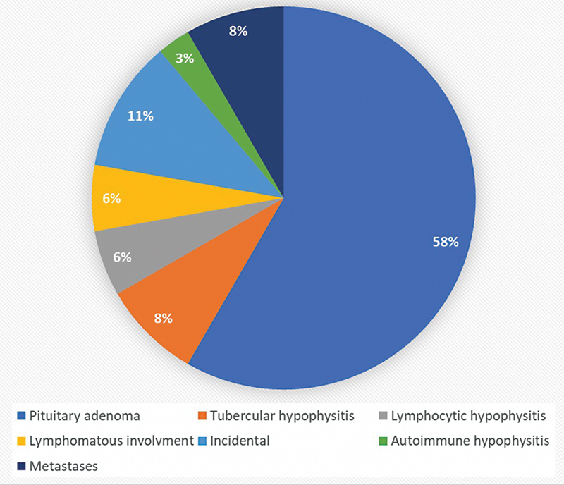
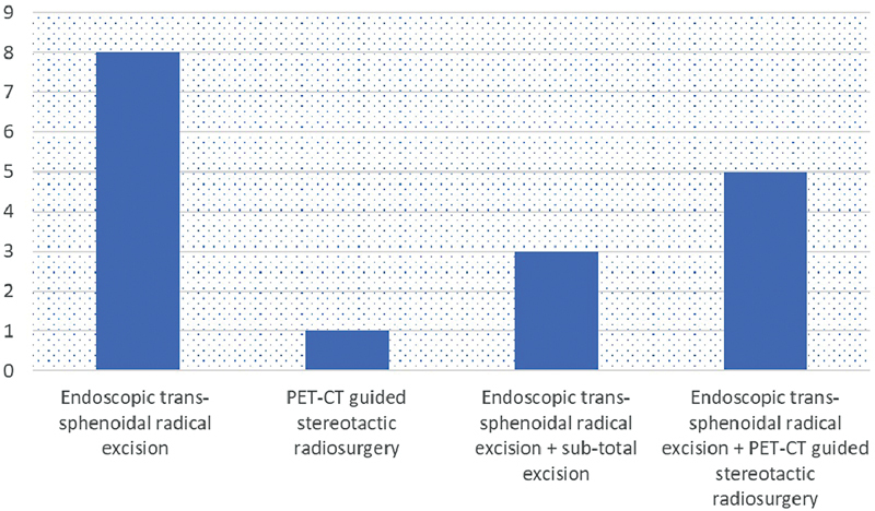
Introduction Pituitary adenoma is the most common disease that affects the gland and may be classified as functional/nonsecretory tumors. Inflammatory/infective causes may also affect the pituitary gland. The 18F-fluorodeoxyglucose positron emission tomography/computed tomography (F18-FDG PET/CT) may have an incremental value in assessing these lesions and in determining their clinical significance. Aim This article assesses the utility of F18-FDG PET/CT in detecting and determining clinical profile of pituitary lesions with abnormal uptake. Methodology Retrospective analysis of all patients who underwent F18-FDG PET/CT from January 2015 to January 2023 was done. Those with abnormal pituitary uptake (standardized uptake value [SUV] > 2.5) were included in the study. SUV value along with relevant anatomical details, biochemical parameters, histopathological details, and follow-up imaging were analyzed. Results Among 15,085 studies, a total of 36 patients (21 males/15 females, average age 47.36 years, range: 17-75 years) with pituitary uptake (0.23%) were included. Out of 36 patients, causes are primary pituitary tumor (21/36, 58%), tubercular hypophysitis (3/36, 8%), lymphocytic hypophysitis (2/36, 6%), lymphomatous involvement (2/36, 6%), autoimmune hypophysitis (1/36, 3%), questionable significance/incidental (4/36, 11%), and metastasis (3/36, 8%)-one each from neuroendocrine tumor ileum, chondrosarcoma, and adenocarcinoma lung. There was no difference in the SUV range between the different etiologies. Among 21 patients with pituitary tumor, biochemical evaluation was done in 19 patients. Two patients were lost to follow-up and did not have biochemical evaluation. Among them, 8 underwent endoscopic transsphenoidal radical excision and 1 patient had PET-CT-guided stereotactic radiosurgery alone. In another 8 patients who had prior endoscopic transsphenoidal radical excision, uptake was noted as residual lesion on PET-CT. Of them, 3 underwent subtotal excision and 5 had PET-CT-guided stereotactic radiosurgery. Biopsy was done in 14 patients, of which 11 were macroadenoma and 3 were microadenoma. Overall, magnetic resonance imaging (MRI) brain was performed in 22 of them and the findings were concordant with F18-FDG PET/CT. Conclusion F18-FDG PET/CT is a useful modality in the evaluation of pituitary uptake. It has an incremental value along with MRI brain and biochemical parameters and is useful for follow-up. Due to its high diagnostic accuracy, it is particularly useful in those with suspected residual/recurrent adenomas.


 分享
分享
 求助内容:
求助内容: 应助结果提醒方式:
 扫码关注我们
