{"title":"骨髓以外:用18f - fdg PET/CT揭示ALL复发的不常见部位。","authors":"Siven Kar, Harshita Gupta, Nusrat Shaikh, Vikram Lele","doi":"10.1055/s-0044-1787894","DOIUrl":null,"url":null,"abstract":"<p><p>Extramedullary infiltration of acute lymphoblastic leukemia/lymphoma (ALL) to genital organs is extremely rare. Here, we present a case report of an asymptomatic 49-year-old female, known case of precursor B-cell ALL, who was incidentally detected with thickened and heterogeneously hyperechoic endometrium on sonography. Contrast magnetic resonance imaging detected large polypoidal enhancing lesions showing intense diffusion restriction occupying the endometrial cavity and similar lesions in the left adnexa, left ovary, and fallopian tube which were suspicious for leukemic infiltration because of the clinical history and atypical appearance of the lesions. <sup>18</sup> F-fluorodeoxyglucose positron emission tomography/computed tomography ( <sup>18</sup> F-FDG PET/CT) was done which revealed intensely metabolically active lesion in the endometrial cavity, left adnexa, omental nodules, retroperitoneal lymph node, pancreatic lesion, and few irregular nodules in the right lower lobe. Biopsy findings confirmed extramedullary relapse of ALL. Hence, <sup>18</sup> F-FDG PET/CT can act as a good whole body survey to look for extramedullary sites of relapse.</p>","PeriodicalId":23742,"journal":{"name":"World Journal of Nuclear Medicine","volume":"23 4","pages":"282-284"},"PeriodicalIF":0.9000,"publicationDate":"2024-06-24","publicationTypes":"Journal Article","fieldsOfStudy":null,"isOpenAccess":false,"openAccessPdf":"https://www.ncbi.nlm.nih.gov/pmc/articles/PMC11637637/pdf/","citationCount":"0","resultStr":"{\"title\":\"Beyond the Marrow: Unveiling Uncommon Sites of ALL Relapse with <sup>18</sup> F-FDG PET/CT.\",\"authors\":\"Siven Kar, Harshita Gupta, Nusrat Shaikh, Vikram Lele\",\"doi\":\"10.1055/s-0044-1787894\",\"DOIUrl\":null,\"url\":null,\"abstract\":\"<p><p>Extramedullary infiltration of acute lymphoblastic leukemia/lymphoma (ALL) to genital organs is extremely rare. Here, we present a case report of an asymptomatic 49-year-old female, known case of precursor B-cell ALL, who was incidentally detected with thickened and heterogeneously hyperechoic endometrium on sonography. Contrast magnetic resonance imaging detected large polypoidal enhancing lesions showing intense diffusion restriction occupying the endometrial cavity and similar lesions in the left adnexa, left ovary, and fallopian tube which were suspicious for leukemic infiltration because of the clinical history and atypical appearance of the lesions. <sup>18</sup> F-fluorodeoxyglucose positron emission tomography/computed tomography ( <sup>18</sup> F-FDG PET/CT) was done which revealed intensely metabolically active lesion in the endometrial cavity, left adnexa, omental nodules, retroperitoneal lymph node, pancreatic lesion, and few irregular nodules in the right lower lobe. Biopsy findings confirmed extramedullary relapse of ALL. Hence, <sup>18</sup> F-FDG PET/CT can act as a good whole body survey to look for extramedullary sites of relapse.</p>\",\"PeriodicalId\":23742,\"journal\":{\"name\":\"World Journal of Nuclear Medicine\",\"volume\":\"23 4\",\"pages\":\"282-284\"},\"PeriodicalIF\":0.9000,\"publicationDate\":\"2024-06-24\",\"publicationTypes\":\"Journal Article\",\"fieldsOfStudy\":null,\"isOpenAccess\":false,\"openAccessPdf\":\"https://www.ncbi.nlm.nih.gov/pmc/articles/PMC11637637/pdf/\",\"citationCount\":\"0\",\"resultStr\":null,\"platform\":\"Semanticscholar\",\"paperid\":null,\"PeriodicalName\":\"World Journal of Nuclear Medicine\",\"FirstCategoryId\":\"1085\",\"ListUrlMain\":\"https://doi.org/10.1055/s-0044-1787894\",\"RegionNum\":0,\"RegionCategory\":null,\"ArticlePicture\":[],\"TitleCN\":null,\"AbstractTextCN\":null,\"PMCID\":null,\"EPubDate\":\"2024/12/1 0:00:00\",\"PubModel\":\"eCollection\",\"JCR\":\"Q4\",\"JCRName\":\"RADIOLOGY, NUCLEAR MEDICINE & MEDICAL IMAGING\",\"Score\":null,\"Total\":0}","platform":"Semanticscholar","paperid":null,"PeriodicalName":"World Journal of Nuclear Medicine","FirstCategoryId":"1085","ListUrlMain":"https://doi.org/10.1055/s-0044-1787894","RegionNum":0,"RegionCategory":null,"ArticlePicture":[],"TitleCN":null,"AbstractTextCN":null,"PMCID":null,"EPubDate":"2024/12/1 0:00:00","PubModel":"eCollection","JCR":"Q4","JCRName":"RADIOLOGY, NUCLEAR MEDICINE & MEDICAL IMAGING","Score":null,"Total":0}
引用次数: 0
摘要
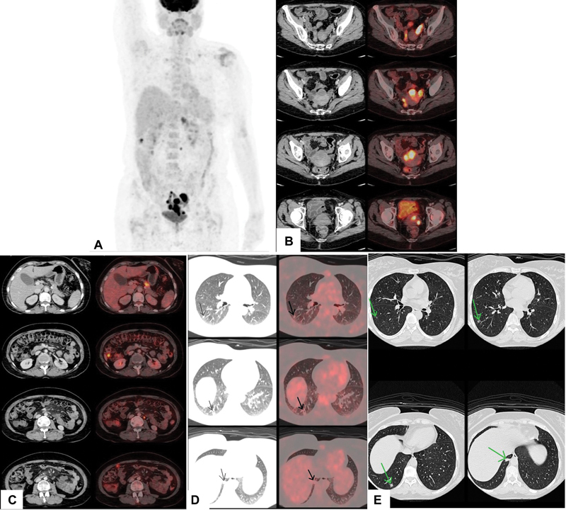
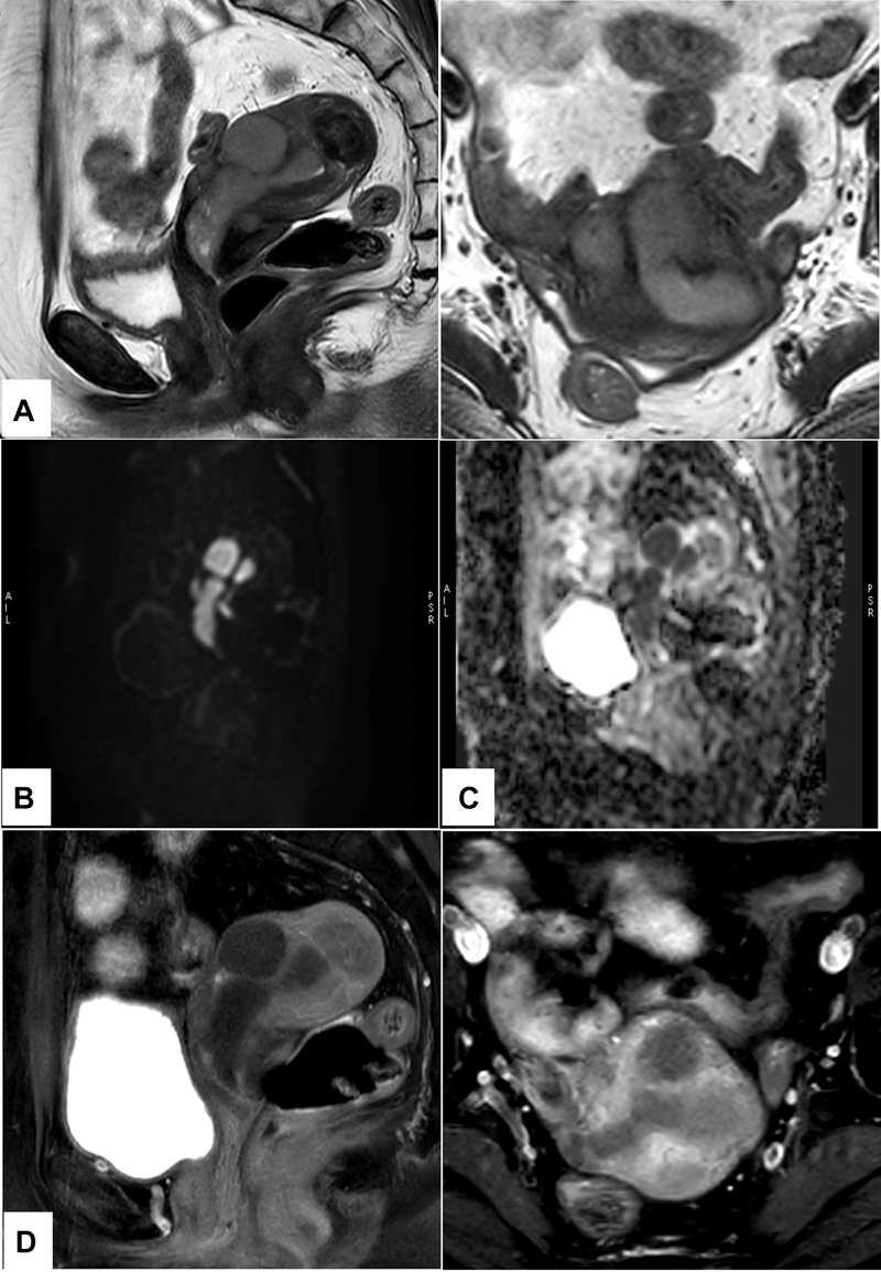
急性淋巴细胞白血病/淋巴瘤(ALL)髓外浸润到生殖器官是非常罕见的。在这里,我们报告一例无症状的49岁女性,已知的前体b细胞ALL病例,偶然发现超声增厚和异质高回声子宫内膜。磁共振造影示大息肉样增强病灶,弥散受限,占据子宫内膜腔,左侧附件、左侧卵巢、输卵管均见类似病灶,临床病史及外观不典型,怀疑白血病浸润。18 f -氟脱氧葡萄糖正电子发射断层扫描/计算机断层扫描(18 F-FDG PET/CT)显示子宫内膜腔、左附件、大网膜结节、腹膜后淋巴结、胰腺病变强烈代谢活跃,右下叶少量不规则结节。活检结果证实ALL髓外复发。因此,18f - fdg PET/CT可以作为一个很好的全身检查来寻找髓外复发部位。
Beyond the Marrow: Unveiling Uncommon Sites of ALL Relapse with 18 F-FDG PET/CT.
Extramedullary infiltration of acute lymphoblastic leukemia/lymphoma (ALL) to genital organs is extremely rare. Here, we present a case report of an asymptomatic 49-year-old female, known case of precursor B-cell ALL, who was incidentally detected with thickened and heterogeneously hyperechoic endometrium on sonography. Contrast magnetic resonance imaging detected large polypoidal enhancing lesions showing intense diffusion restriction occupying the endometrial cavity and similar lesions in the left adnexa, left ovary, and fallopian tube which were suspicious for leukemic infiltration because of the clinical history and atypical appearance of the lesions. 18 F-fluorodeoxyglucose positron emission tomography/computed tomography ( 18 F-FDG PET/CT) was done which revealed intensely metabolically active lesion in the endometrial cavity, left adnexa, omental nodules, retroperitoneal lymph node, pancreatic lesion, and few irregular nodules in the right lower lobe. Biopsy findings confirmed extramedullary relapse of ALL. Hence, 18 F-FDG PET/CT can act as a good whole body survey to look for extramedullary sites of relapse.


 分享
分享
 求助内容:
求助内容: 应助结果提醒方式:
 扫码关注我们
